. 2006 Nov 15;400(1):13-22.
doi: 10.1042/BJ20060664.

Structural and kinetic analyses of the H121A mutant of cholesterol oxidase

Affiliations

Structural and kinetic analyses of the H121A mutant of cholesterol oxidase

Louis Lim et al. Biochem J. .

Abstract

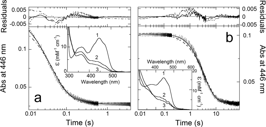
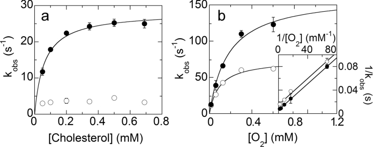
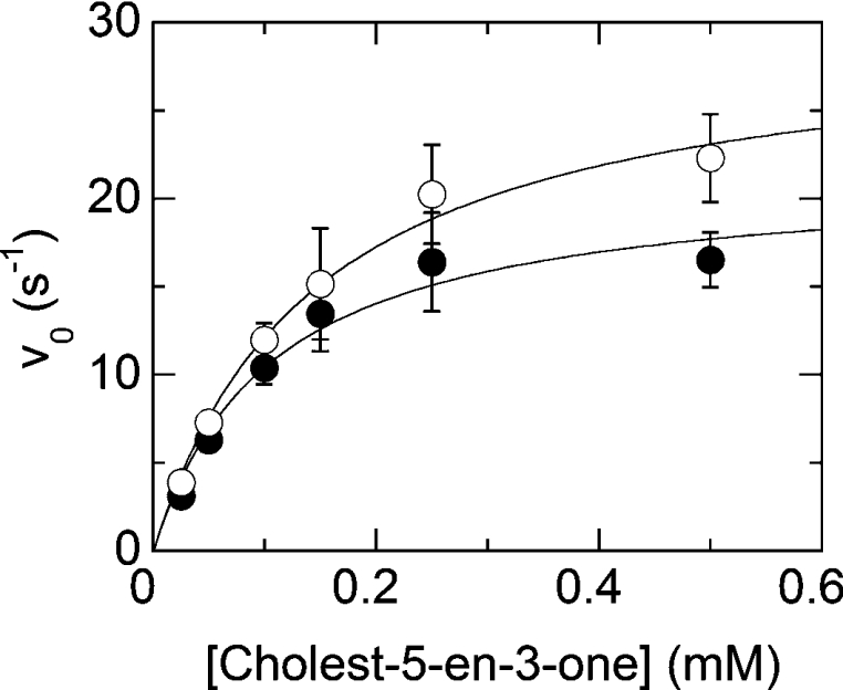
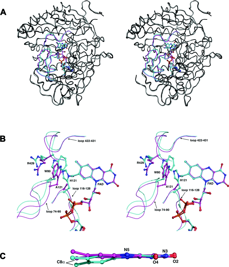
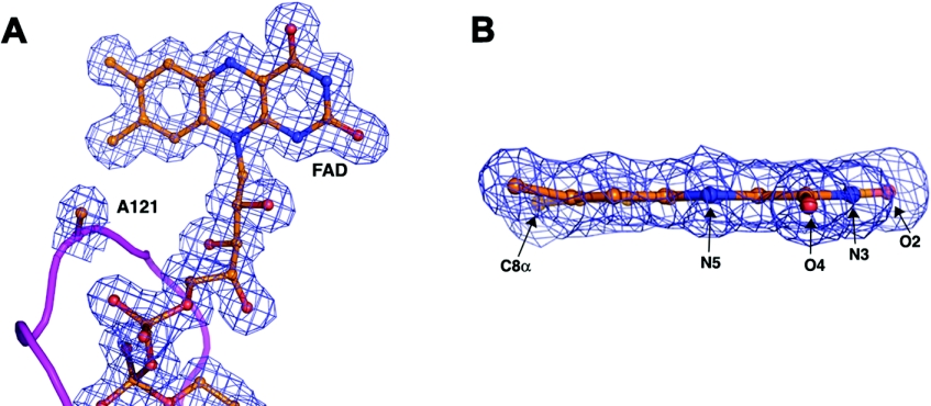
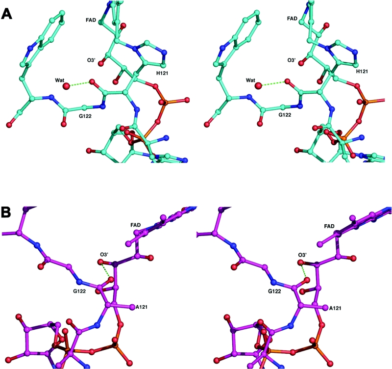
Cholesterol oxidase is a monomeric flavoenzyme that catalyses the oxidation of cholesterol to cholest-5-en-3-one followed by isomerization to cholest-4-en-3-one. The enzyme from Brevibacterium sterolicum contains the FAD cofactor covalently bound to His121. It was previously demonstrated that the H121A substitution results in a approximately 100 mV decrease in the midpoint redox potential and a approximately 40-fold decrease in turnover number compared to wild-type enzyme [Motteran, Pilone, Molla, Ghisla and Pollegioni (2001) Journal of Biological Chemistry 276, 18024-18030]. A detailed kinetic analysis of the H121A mutant enzyme shows that the decrease in turnover number is largely due to a corresponding decrease in the rate constant of flavin reduction, whilst the re-oxidation reaction is only marginally altered and the isomerization reaction is not affected by the substitution and precedes product dissociation. The X-ray structure of the mutant protein, determined to 1.7 A resolution (1 A identical with 0.1 nm), reveals only minor changes in the overall fold of the protein, namely: two loops have slight movements and a tryptophan residue changes conformation by a rotation of 180 degrees about chi1 compared to the native enzyme. Comparison of the isoalloxazine ring moiety of the FAD cofactor between the structures of the native and mutant proteins shows a change from a non-planar to a planar geometry (resulting in a more tetrahedral-like geometry for N5). This change is proposed to be a major factor contributing to the observed alteration in redox potential. Since a similar distortion of the flavin has not been observed in other covalent flavoproteins, it is proposed to represent a specific mode to facilitate flavin reduction in covalent cholesterol oxidase.


Figures

Figure 1
Figure 1. Time courses of the anaerobic reduction of wild-type (a) and H121A (b) BCO
Anaerobic solutions of enzymes (≈9 μM and ≈6.3 μM for wild-type and H121A BCOs respectively) and 0.7 mM cholesterol were mixed in the stopped-flow instrument, in the presence of 1% propan-2-ol/1% Thesit (pH 7.5) at 25 °C; (|) represent the data points at 446 nm. Curve (-−- −-) is the fit for a single exponential decay; curve (---) is the fit for a double exponential decay; curve (−) is the trace obtained from simulations using Specfit/32 software and based on the sequence of steps of Eqn 1, on known extinction coefficients for the oxidized and reduced enzyme forms (see insets) and on the rate constants reported in Table 2. Insets: The spectra shown are those obtained by deconvolution with Specfit/32. Spectrum 1, oxidized enzyme(s); Spectrum 2, reduced enzyme–product intermediate complex; and Spectrum 3, free reduced enzyme. The residuals are the subtraction of the experimental data points at 446 nm from the traces obtained from fit or simulation procedures.
Figure 2
Figure 2. Dependence of the rates of flavin reduction and re-oxidation for wild-type and mutant enzymes
(a) Dependence of the observed rates of flavin reduction for wild-type BCO from the cholesterol concentration. The observed rate constants are obtained from fits of the absorbance change at 446 nm using a double exponential algorithm as detailed in the text (conditions as detailed in the legend of Figure 1). The data points are: (●) the values of kobs1, the rates corresponding to the transformation of species (1) into species (2) (inset of Figure 1a); (○) the values of kobs2, the rates corresponding to the transformation of species (2) into species (3) (inset of Figure 1a). Where error bars are not shown the data scatter is smaller than the size of the symbols. (b) Dependence of the rates of flavin re-oxidation for wild-type (●) and H121A BCO (○) from the oxygen concentration. The observed rate constants were obtained from fits of traces reflecting the absorbance increase at 446 nm (the time courses are essentially monophasic). Where error bars are not shown the data scatter is smaller than the size of the symbols. Inset: double reciprocal plot of the same data as reported in the main graph.
Figure 3
Figure 3. Dependence of the observed rate of isomerization of cholest-5-en-3-one into cholest-5-en-4-one from the substrate concentration for the oxidized (●) and reduced (○) forms of H121A BCO
The data points (vo) are the rates of the absorbance changes at 240 nm that reflect isomerization as obtained in stopped-flow experiments (see text for details). Where error bars are not shown the data scatter is smaller than the size of the symbols.
Figure 4
Figure 4. Comparison of the structures of wild-type and H121A BCO
(a) Stereo figure showing a superposition of the trace of wild-type BCO and the H121A mutant enzyme. The three loops that exhibit the largest change between the two structures are coloured in magenta (for the mutant BCO) and cyan (for the wild-type BCO). The remaining structural elements are coloured in light (mutant enzyme) and dark (wild-type BCO) grey. The FAD molecules are coloured in the same way as the loops of the corresponding structure. The superposition was carried out using the SSM algorithm [46] incorporated into COOT [47]. (b). Stereo view of key residues in the loop regions that have altered positions in the native and mutant enzyme structure. The colouring is as detailed above. The side-chains for His121, Ala121 (mutant BCO), Trp80 and Arg429 for both structures are included. (c) Side view of the superposed isoalloxazine moiety for the native enzyme (cyan) and the H121A mutant BCO (magenta). The atoms in the pyrimidine ring were used for the superposition.
Figure 5
Figure 5. The 2 FoFc electron density map for the H121A mutant protein
The map, contoured at 1.5 σ, shows density features for (a) the FAD cofactor and residue 121 and (b) a side view of the isoalloxazine ring moiety.
Figure 6
Figure 6. Interactions in the region of residues 121 and 122
Stereo view showing the intramolecular interactions in the region of 121–122 for (a) the native enzyme and (b) the mutant BCO. Hydrogen bonds are depicted as green broken lines.

Similar articles

Cited by

References

    1. MacLachlan J., Wotherspoon A. T. L., Ansell R. O., Brooks C. J. W. Cholesterol oxidase: sources, physical properties and analytical applications. J. Steroid Biochem. Mol. Biol. 2000;72:169–195. - PubMed
    1. Corbin D. R., Greenplate J. T., Purcell J. P. The identification and development of proteins for control of insects in genetically modified crops. HortScience. 1998;33:614–617.
    1. Navas J., Gonzalez-Zorn B., Ladron N., Garrido P., Vazquez-Boland J. A. Identification and mutagenesis by allelic exchange of choE, encoding a cholesterol oxidase from the intracellular pathogen Rhodococcus equi. J. Bacteriol. 2001;183:4796–4805. - PMC - PubMed
    1. Li J., Vrielink A., Brick P., Blow D. M. Crystal structure of cholesterol oxidase complexed with a steroid substrate: Implications for flavin adenine dinucleotide dependent alcohol oxidases. Biochemistry. 1993;32:11507–11515. - PubMed
    1. Ladron N., Fernandez M., Aguero J., Gonzalez Zorn B., Vazquez-Boland J. A., Navas J. Rapid identification of Rhodococcus equi by a PCR assay targeting the choE gene. J. Clin. Microbiol. 2003;41:3241–3245. - PMC - PubMed

Publication types

MeSH terms

Associated data