. 2006 Jul 21;23(2):231-9.
doi: 10.1016/j.molcel.2006.06.023.

Pulling on the nascent RNA during transcription does not alter kinetics of elongation or ubiquitous pausing

Affiliations

Pulling on the nascent RNA during transcription does not alter kinetics of elongation or ubiquitous pausing

Ravindra V Dalal et al. Mol Cell. .

Abstract

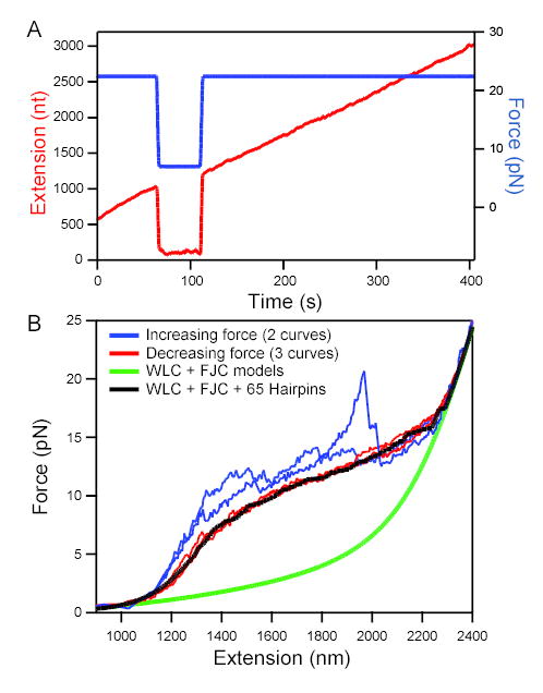
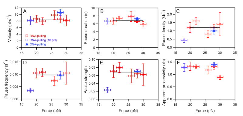
Transcriptional elongation and termination by RNA polymerase (RNAP) are controlled by interactions among the nascent RNA, DNA, and RNAP that comprise the ternary transcription elongation complex (TEC). To probe the effects of cotranscriptionally folded RNA hairpins on elongation as well as the stability of the TEC, we developed a single-molecule assay to monitor RNA elongation by Escherichia coli RNAP molecules while applying controlled loads to the nascent RNA that favor forward translocation. Remarkably, forces up to 30 pN, twice those required to disrupt RNA secondary structure, did not significantly affect enzyme processivity, transcription elongation rates, pause frequencies, or pause lifetimes. These results indicate that ubiquitous transcriptional pausing is not a consequence of the formation of hairpins in the nascent RNA. The ability of the TEC to sustain large loads on the transcript reflects a tight binding of RNA within the TEC and has important implications for models of transcriptional termination.


Figures

Figure 1
Figure 1. Cartoon of experimental assays and data records (not to scale)
(A) The RNA-pulling assay. RNAP (green) transcribing a DNA template (blue) is attached to a bead via an avidin-biotin linkage (yellow/black). Nascent RNA (red) from the polymerase is hybridized to a 3-kb-long DNA handle (green) via a 25-base overhang; the distal end of the handle is attached via a digoxigenin-antibody linkage (black/purple) to the coverglass of a flow cell mounted on a 3D piezo stage. The optical trap (pink) holds the bead at a fixed offset from the trap center, producing a constant restoring force. The stage is moved by feedback to compensate for any elongation of the tether. (B) The DNA-pulling assay for assisting load. The upstream end of the DNA template is attached to the coverglass surface via a digoxigenin-antibody linkage; the nascent RNA remains unbound and free to form secondary structure. (C) 9 representative transcription records (of N = 202) from the RNA-pulling assay (red), showing elongation of the nascent RNA (in nt) vs. time. Note transcriptional pauses. (D) 6 representative transcription records (of N = 87) from the DNA-pulling assay (blue), showing progress along the DNA (in bp) vs. time. Note transcriptional pauses.
Figure 2
Figure 2. Secondary structure in the nascent RNA is suppressed at high force
(A) RNA extension (red; left axis) vs. time at high and low loads. Elongation took place at an initial force of 22 pN (blue; right axis). When force was reduced to 7 pN at 75 s, apparent elongation ceased. When high force was restored at 110 s, extension returned to the position extrapolated from earlier elongation, showing that transcription had continued during the low-force period with no significant change in extension as secondary structure formed. (B) Force-extension curves (FECs) for a tether that had stalled prematurely (taken at ~120 nm/s). Stretching curves (increasing force; 2 examples shown) displayed distinct features with each pull (blue traces). In contrast, relaxation curves (decreasing force; 3 examples shown) displayed very reproducible behavior (red traces). Model FECs: A worm-like chain (WLC, representing dsDNA) plus a freely-jointed chain (FJC, fit to 2,200 nt ssRNA) fits the data only in low and high force limits (green line). The additional effect of forming 65 random hairpins (loop 4 nt; Gaussian distribution of stem lengths centered at 10.5 bp, std. dev. 3 bp) reproduces the plateau seen in the experimental data (black line) (see Supplemental Material).
Figure 3
Figure 3. Normalized pause lifetime distributions for RNA-pulling (red) and DNA-pulling (blue) assays at 26–30 pN load
Lifetime distributions were scaled by total number of seconds transcribed (RNA: t = 14,608 s, N = 132; DNA: tt = 12,042 s, N = 122) to supply the overall frequency (pauses/s). Bin widths were ≥1 sec and scaled to ensure ≥6 counts per bin; statistical errors were computed from √N. Inset: Semi-logarithmic plot for DNA (filled triangles) and RNA (open squares). Fits to DNA data: Double exponential τ = 0.9 ± 0.4 and 5.3 ± 1.6 s (χν2 = 0.81; ν= 3; pν2) = 0.48; 4 parameters), single exponential (not shown) τ = 2.7 ± 0.3 (χν2 = 3.1; ν = 5; pν2) = 0.009; 2 parameters). Fits to RNA data: Double exponential τ = 0.6 ± 0.2 and 3.8 ± 0.2 s (χν2 = 0.99; ν = 3; pν2) = 0.39; 4 parameters); single exponential (not shown) τ = 2.4 ± 0.3 (χν2 = 3.31; ν = 5; pν2) = 0.003; 2 parameters).
Figure 4
Figure 4. Force dependence of velocity, pause characteristics, and processivity
RNA-pulling data (open squares), DNA-pulling data (filled triangles). Fits to weighted means (black horizontal lines) rather than to lines (nonzero slope) were justified by F tests. The low force point at 18 pN was excluded from fits because it was deemed too close to the opening force for the most stable hairpins. Estimated errors represent std. errs. in (A, B) and bootstrap errors in (C-F). (A) Mean velocity vs. force; avg. = 8.6 ± 0.7 nt/s. (B) Mean pause duration vs. force; avg. = 6.9 ± 0.2 s. (C) Pause density vs. force; avg. = 1.2 ± 0.1 kb−1. (D) Pause frequency vs. force; avg. = 9.7 ± 0.9 ×10⁻³ s−1. (E) Pause strength (pause duration multiplied by frequency) vs. force; avg. = 68 ± 7 ×10⁻³. (F) Apparent processivity (distance to tether rupture) vs. force.

Similar articles

Cited by

References

    1. Abbondanzieri EA, Greenleaf WJ, Shaevitz JW, Landick R, Block SM. Direct observation of base-pair stepping by RNA polymerase. Nature. 2005;438:460–465. - PMC - PubMed
    1. Adelman K, La Porta A, Santangelo TJ, Lis JT, Roberts JW, Wang MD. Single molecule analysis of RNA polymerase elongation reveals uniform kinetic behavior. Proc Natl Acad Sci U S A. 2002;99:13538–13543. - PMC - PubMed
    1. Adelman K, Yuzenkova J, La Porta A, Zenkin N, Lee J, Lis JT, Borukhov S, Wang MD, Severinov K. Molecular mechanism of transcription inhibition by Peptide antibiotic microcin j25. Mol Cell. 2004;14:753–762. - PubMed
    1. Artsimovitch I, Landick R. Pausing by bacterial RNA polymerase is mediated by mechanistically distinct classes of signals. Proc Natl Acad Sci U S A. 2000;97:7090–7095. - PMC - PubMed
    1. Artsimovitch I, Landick R. The transcriptional regulator RfaH stimulates RNA chain synthesis after recruitment to elongation complexes by the exposed nontemplate DNA strand. Cell. 2002;109:193–203. - PubMed

Publication types