. 2006 Oct 1;576(Pt 1):135-49.
doi: 10.1113/jphysiol.2006.115246. Epub 2006 Jul 20.

Regulation of the desensitization and ion selectivity of ATP-gated P2X2 channels by phosphoinositides

Affiliations

Regulation of the desensitization and ion selectivity of ATP-gated P2X2 channels by phosphoinositides

Yuichiro Fujiwara et al. J Physiol. .

Abstract

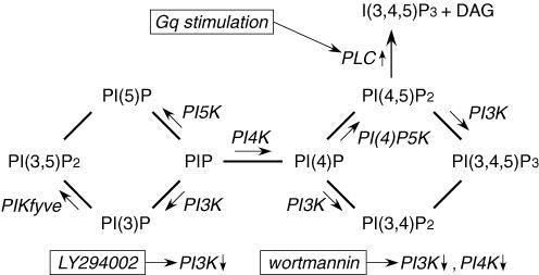
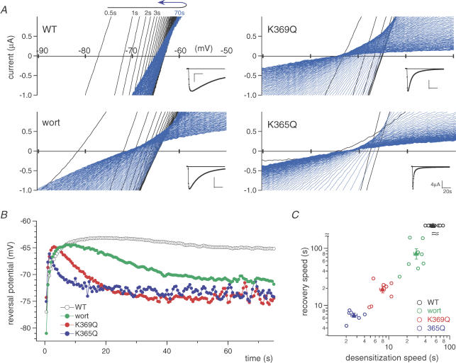
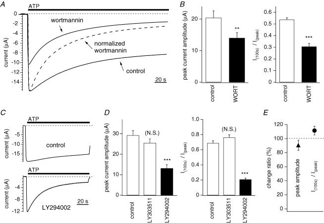
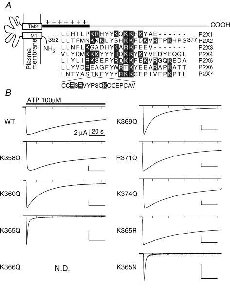
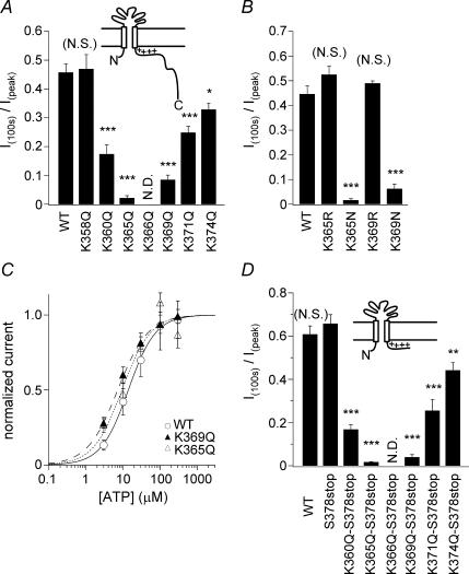
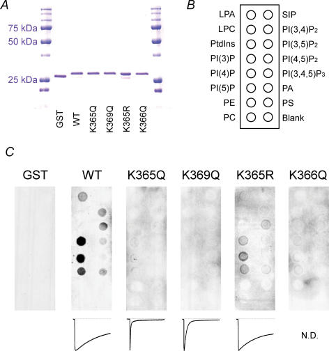
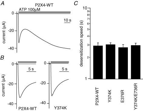
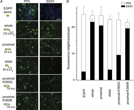
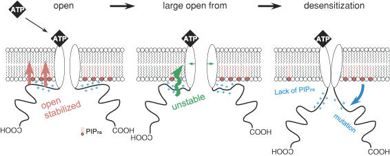
Phosphoinositides (PIP(n)s) are known to regulate the activity of some ion channels. Here we determined that ATP-gated P2X(2) channels also are regulated by PIP(n)s, and investigated the structural background and the unique features of this regulation. We initially used two-electrode voltage clamp to analyse the electrophysiological properties of P2X(2) channels expressed in Xenopus oocytes, and observed that preincubation with wortmannin or LY294002, two PI3K inhibitors, accelerated channel desensitization. K365Q or K369Q mutation of the conserved, positively charged, amino acid residues in the proximal region of the cytoplasmic C-terminal domain also accelerated desensitization, whereas a K365R or K369R mutation did not. We observed that the permeability of the channel to N-methyl-d-glucamine (NMDG) transiently increased and then decreased after ATP application, and that the speed of the decrease was accelerated by K365Q or K369Q mutation or PI3K inhibition. Using GST-tagged recombinant proteins spanning the proximal C-terminal region, we then analysed their binding of the P2X(2) cytoplasmic domain to anionic lipids using PIP(n)s-coated nitrocellulose membranes. We found that the recombinant proteins that included the positively charged region bound to PIPs and PIP(2)s, and that this binding was eliminated by the K365Q and K369Q mutations. We also used a fluorescence assay to confirm that fusion proteins comprising the proximal C-terminal region of P2X(2) with EGFP expressed in COS-7 cells closely associated with the membrane. Taken together, these results show that membrane-bound PIP(n)s play a key role in maintaining channel activity and regulating pore dilation through electrostatic interaction with the proximal region of the P2X(2) cytoplasmic C-terminal domain.


Figures

Figure 1
Figure 1. Metabolic map showing the relationships among various PIPns and PI-kinases
The sites of action of the indicated PI-kinases, kinase inhibitors and Gq-coupled receptor stimulation are indicated.
Figure 6
Figure 6. The relationship between channel desensitization and the time-dependent changes in the permeability to NMDG
A, representative time-dependent changes in the current–voltage relationships in NMDG-based solution after ATP application. Recordings were obtained by applying ramp pulses every 0.5 s to oocytes expressing WT or mutant P2X2 receptors. The inset shows current traces measured at −90 mV. In each recording, shifts in the direction of depolarization are shown in black, while shifts in the direction of hyperpolarization (recovery) are shown in blue. B, time-dependent shifts in the reversal potentials derived from the data in A. The symbols used are as indicated in the figure, wort = wortmannin. C, relationship between the speed of recovery of the shift in the reversal potential and the speed of the reduction in current amplitude (desensitization) in NMDG-based external solution. The time constants of the recovery are plotted against those of the desensitization for each of the oocytes recorded (C, open circles). Filled triangles and bars depict means ± s.e.m. of each group (n = 11 (WT), n = 8 (wort), n = 8 (K369Q) and n = 6 (K365Q)).
Figure 2
Figure 2. Effect of PI3K inhibitors and Gq-coupled receptor stimulation on desensitization of P2X2 receptor channel
A and C, currents carried by WT P2X2 channels expressed in Xenopus oocytes were recorded using two-electrode voltage clamp in Na+-based external solution. The holding potential was −60 mV, and 100 μm ATP was applied. B, D and E, statistical comparison of the peak current amplitudes and the extent of desensitization. The ratios of the current amplitudes measured 100 s after the application of ATP and the peak amplitudes (I100s/Ipeak) were calculated as an index of desensitization. A, representative currents recorded from oocytes with or without preincubation for 2 h with 30 μm wortmannin. Also shown is the normalized current trace obtained with wortmannin. B, comparison of the data in A. Bars depict means ± s.e.m. (n = 10); means were compared statistically using Student's t test (***P < 0.001, **P < 0.01). C, representative currents recorded with and without preincubation for 12 h with 30 μm LY294002. D, comparison of the data in C. Bars depict means ± s.e.m. (n = 7–8); means were compared statistically using Tukey's test (***P < 0.001 (N.S.): P > 0.005). E, Peak amplitudes (▴) and I100s/Ipeak ratios (○) before and after stimulation with 1 μm substance P for 10 min in cells coexpressing P2X2 channels and substance P receptors. Bars depict means ± s.e.m. (n = 11).
Figure 3
Figure 3. The effect of substituting the positively charged amino acid residues in the proximal region of the cytoplasmic C-terminal domain of P2X2on desensitization
A, alignment of the amino acid sequences of the proximal C-terminal regions of P2X family proteins. The original reports used for the alignment are as follows: P2X1 (Valera et al. 1994), P2X2 (Brake et al. 1994), P2X3 (Lewis et al. 1995), P2X4 (Seguela et al. 1996), P2X5 (Collo et al. 1996), P2X6 (Collo et al. 1996), P2X7 (Surprenant et al. 1996). Positively charged residues (lysine and arginine) are highlighted. B, representative macroscopic currents of WT and mutant P2X2 channels recorded as in Fig. 2. N.D. denotes a channel that did not show detectable currents, and so could not be analyzed.
Figure 4
Figure 4. Analysis of the effects of the indicated mutations on the extent of P2X2 channel desensitization
A, B and D, comparison of the extent in desensitization of WT P2X2 and mutant channels. Currents were recorded as in Fig. 2. Bars depict means ± s.e.m. (n = 5–7 in A, n = 7 in B and n = 5–10 in D); control means (control= WT in A, control = WT in B and control = S378stop in D) and those of each mutant were compared statistically using Tukey's test (***P < 0.001, **P < 0.01, *P < 0.05 and (N.S) means P > 0.05). C, concentration–response relationships of WT P2X2 and the indicated mutants.
Figure 7
Figure 7. Binding of the proximal region of the P2X2 cytoplasmic C-terminal domain to phospholipids
A, recombinant GST-tagged WT and mutant proximal C-terminal regions of P2X2 were resolved by SDS-PAGE and stained with Coomassie Brilliant Blue. B, positions of the indicated phospholipids blotted on a nitrocellulose membrane. Abbreviations used are as follows: Lyso phosphatidic acid (LPA); lysophosphocholine (LPC); sphingosine-1-phosphate (SIP); phosphatidic acid (PA); phosphatidyl choline (PC); phosphatidyl serine (PS); phosphatidyl ethanolamine (PE). C, binding patterns of the WT and K365Q, K369Q, K365R K366Q mutant proteins and a negative control protein (GST) to phospholipids blotted on the membrane.
Figure 5
Figure 5. Effects on desensitization of introducing positively charged amino acid residues into the C-terminal domain of P2X4
A, representative current through the WT P2X4 channel. An initial fast desensitization phase and a later slow resensitization phase were observed. B, expanded recordings of the desensitization phase of WT P2X4 and the Y374K mutant. C, comparison of the speed of desensitization of WT and mutant P2X2 channels. I100s/Ipeak was not usable as an index here, so the desensitization phase was fitted with a single exponential function, and the time constants were determined. Bars depict means ± s.e.m. (n = 7).
Figure 8
Figure 8. Interaction of the P2X2 cytoplasmic C-terminal domain with the plasma membrane
A, expression of EGFP and EGFP-P2X2 fusion proteins in transiently transfected COS-7 cells. The abbreviations used are as follows: EGFP alone, EGFP protein used as a negative control; whole, EGFP with the entire cytoplasmic C-terminus (L353–L472); proximal, EGFP with the proximal C-terminal region (L353–S378stop); distal, EGFP with the distal C-terminal region (S378–L472); proximal K365Q or K365R, EGFP with the proximal C-terminus carrying the K365Q or K365R mutation, respectively. Cells were fixed in either paraformaldehyde or ethanol. Images were acquired with a 20× objective lens and an optical filter set for EGFP. In the images of EGFP alone, DAPI staining also is shown to confirm that there are sufficient numbers of cells in the visual field. The cell density is similar in the other images, but DAPI staining was omitted for clarity. B, quantitative comparison of the intensity of the fluorescence signals obtained with the indicated five constructs.
Figure 9
Figure 9. Schematic diagram explaining the membrane PIPn-dependent conformational changes in the P2X2 channel
For convenience, only the cytoplasmic C-terminal domain and the transmembrane pore region of the channel are shown; omitted are the extracellular domain and the cytoplasmic N-terminal region. Open: an initial open state with a low permeability to NMDG. Large open form: a state showing a high permeability to NMDG. The equilibrium is inclined toward the desensitized state when interaction with the plasma membrane is not maintained. Conformational changes in the proximal cytoplasmic region are thought to occur during transitions from the open to the large open state, and from large open to the desensitized state.

References

    1. Baukrowitz T, Schulte U, Oliver D, Herlitze S, Krauter T, Tucker SJ, Ruppersberg JP, Fakler B. PIP2 and PIP as determinants for ATP inhibition of KATP channels. Science. 1998;282:1141–1144. - PubMed
    1. Bichet D, Lin YF, Ibarra CA, Huang CS, Yi BA, Jan YN, Jan LY. Evolving potassium channels by means of yeast selection reveals structural elements important for selectivity. Proc Natl Acad Sci U S A. 2004;101:4441–4446. - PMC - PubMed
    1. Brake AJ, Wagenbach MJ, Julius D. New structural motif for ligand-gated ion channels defined by an ionotropic ATP receptor. Nature. 1994;371:519–523. - PubMed
    1. Chaumont S, Jiang LH, Penna A, North RA, Rassendren F. Identification of a trafficking motif involved in the stabilization and polarization of P2X receptors. J Biol Chem. 2004;279:29628–29638. - PubMed
    1. Chemin J, Patel AJ, Duprat F, Lauritzen I, Lazdunski M, Honore E. A phospholipid sensor controls mechanogating of the K+ channel TREK-1. EMBO J. 2005;24:44–53. - PMC - PubMed

Publication types

MeSH terms

LinkOut - more resources