Review
. 2006 Oct 15;576(Pt 2):341-7.
doi: 10.1113/jphysiol.2006.110270. Epub 2006 Jul 20.

Can homeostatic circuits learn and remember?

Affiliations
Review

Can homeostatic circuits learn and remember?

Grant R J Gordon et al. J Physiol. .

Abstract

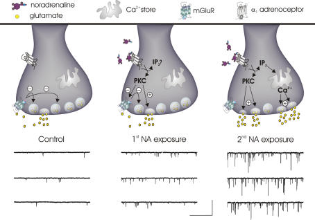
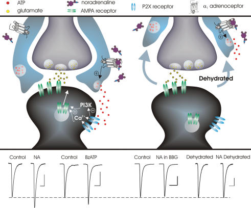
Alterations in synaptic strength are thought to represent the cellular basis of learning and memory. While such processes appear to be fundamental to all synapses, until recently there has been a relative dearth of information regarding synaptic 'memory' processes in autonomic circuits. Here we examine recent advances in our understanding of plasticity at glutamatergic synapses onto magnocellular neurosecretory cells in the hypothalamus, paying particular attention to the contributions of noradrenaline in coding long-lasting pre- and postsynaptic changes in efficacy. We also highlight recent work demonstrating that glial cells play a crucial role in the induction of long-term potentiation. Based on the work reviewed here, we have a clearer picture of the synaptic and cellular mechanisms that allow autonomic pathways to learn and remember.


Figures

Figure 1
Figure 1. Priming of mESPC frequency and amplitude by NA
Left panel: in control conditions, synaptic glutamate activates presynaptic mGluRs keeping mEPSC frequency low. Middle panel: initial α1-adrenoceptor activation increases PKC activity causing an enhancement of mEPSC frequency without changing mEPSC amplitude. Although the facilitating effect on mEPSC frequency is still limited by the activity of functional mGluRs, during this time PKC is working to inactivate these autoreceptors. Right panel: the consequences of mGluR inactivation become apparent when an additional NA challenge is administered in which an increase in mEPSC frequency is observed that is substantially larger than the first. Successive α1-adrenoceptor activation also results in dramatically larger mEPSCs that arise from the rapid release of stored calcium. The mechanism underlying amplitude priming, however, has not been elucidated. The voltage clamp traces of mEPSCs are adapted from Gordon & Bains (2003). Scale bars, 50 pA and 1 s.
Figure 2
Figure 2. Induction of long-term synaptic strengthening by NA-mediated release of glial ATP depends on the physical neuro-glia relationship
Upper panel, left: in the control state, where there is a relative abundance of glial processes surrounding synaptic elements, glial cell α1-adrenoceptor activation triggers the release of ATP which can then activate postsynaptic P2X channels on MNCs. P2X channel activation results in calcium influx and the activation of phosphotidyl inositol 3-kinase (PI3K) leading to the insertion of AMPA receptors which is manifested as a long lasting increase in mEPSC amplitude. Upper panel, right: during states of chronic dehydration or lactation where there is a withdrawal of glial processes from around synaptic elements, NA fails to elicit changes in mEPSC amplitude. Lower panel: average mEPSC traces taken during control and 30 min after treatment. Left: NA causes a long-lasting increase in mEPSC amplitude, an effect that is mimicked by the P2X receptor agonist BzATP. Right: the long-lasting enhancement of mEPSC amplitude caused by NA is blocked either by the P2X7 antagonist Brilliant Blue G (BBG) or by withdrawal of glial processes. Scale bars: 10pA, 5 ms. Data adapted from Gordon et al. (2005).
Figure 3
Figure 3. Long-lasting plasticity at glutamatergic synapses on MNCs
Summary of different types of enduring plasticity at glutamatergic synapses on MNCs, which incorporate presynaptic, postsynaptic and glial signalling to enhance excitatory drive for extended periods of time. NA utilizes the presynaptic terminal to prime glutamate release by inactivating mGluRs and to trigger MVR by recruiting calcium release from internal stores. The two remaining elements of the tripartite synapse are utilized concurrently to elicit AMPA receptor insertion postsynaptically and thus a long-lasting change in synaptic strength. NA acts on glial cells to trigger the release of ATP which subsequently acts on postsynaptic P2X channels to induce activity-independent changes in synapse function. Finally, glia-derived d-serine acts as an essential co-transmitter with synaptically released glutamate to activate postsynaptic NMDA receptors in the induction of classical activity-dependent plasticity.

References

    1. Anderson CM, Bergher JP, Swanson RA. ATP-induced ATP release from astrocytes. J Neurochem. 2004;88:246–256. - PubMed
    1. Armstrong WE, Gallagher MJ, Sladek CD. Noradrenergic stimulation of supraoptic neuronal activity and vasopressin release in vitro: mediation by an alpha 1-receptor. Brain Res. 1986;365:192–197. - PubMed
    1. Bains JS, Longacher JM, Staley KJ. Reciprocal interactions between CA3 network activity and strength of recurrent collateral synapses. Nat Neurosci. 1999;2:720–726. - PubMed
    1. Boudaba C, Linn DM, Halmos KC, Tasker JG. Increased tonic activation of presynaptic metabotropic glutamate receptors in the rat supraoptic nucleus following chronic dehydration. J Physiol. 2003;551:815–823. - PMC - PubMed
    1. Bourque CW, Kirkpatrick K, Jarvis CR. Extrinsic modulation of spike afterpotentials in rat hypothalamoneurohypophysial neurons. Cell Mol Neurobiol. 1998;18:3–12. - PMC - PubMed

Publication types

MeSH terms

Substances

LinkOut - more resources