. 2006 Aug 1;103(31):11689-94.
doi: 10.1073/pnas.0600275103. Epub 2006 Jul 20.

Nitric oxide inhibits exocytosis of cytolytic granules from lymphokine-activated killer cells

Affiliations

Nitric oxide inhibits exocytosis of cytolytic granules from lymphokine-activated killer cells

Marcella Ferlito et al. Proc Natl Acad Sci U S A. .

Abstract

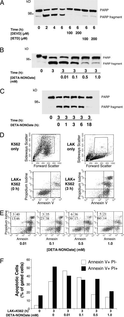
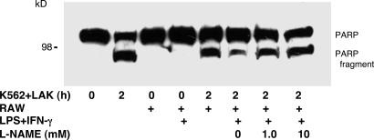
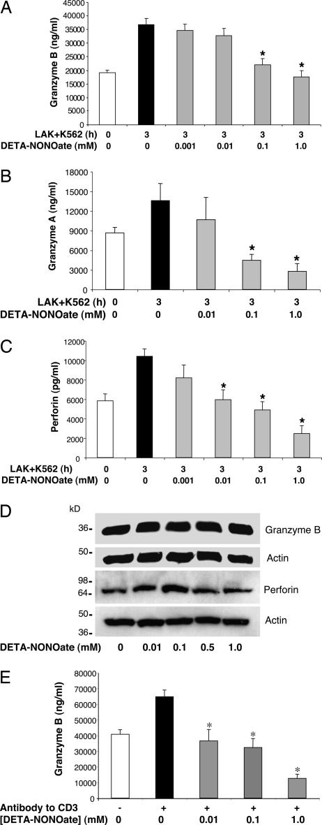
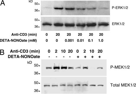
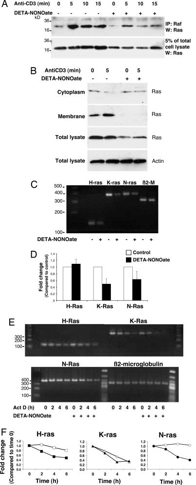
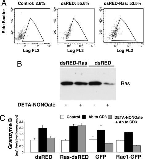
NO inhibits cytotoxic T lymphocyte killing of target cells, although the precise mechanism is unknown. We hypothesized that NO decreases exocytosis of cytotoxic granules from activated lymphocytes. We now show that NO inhibits lymphokine-activated killer cell killing of K562 target cells. Exogenous and endogenous NO decreases the release of granzyme B, granzyme A, and perforin: all contents of cytotoxic granules. NO inhibits the signal transduction cascade initiated by cross-linking of the T cell receptor that leads to granule exocytosis. In particular, we found that NO decreases the expression of Ras, a critical signaling component within the exocytic pathway. Ectopic expression of Ras prevents NO inhibition of exocytosis. Our data suggest that Ras mediates NO inhibition of lymphocyte cytotoxicity and emphasize that alterations in the cellular redox state may regulate the exocytic signaling pathway.


Conflict of interest statement

Conflict of interest statement: No conflicts declared.

Figures

Fig. 1.
Fig. 1.
Exogenous NO inhibits LAK cell killing of K562 target cells. (A) LAK cells activate apoptosis in K562 target cells; measurement of PARP cleavage is shown. LAK cells were incubated with K562 cells for increasing periods of time, and cell lysates were analyzed by Western blotting with antibody to PARP. Some K562 cells were pretreated with inhibitors of apoptosis [DEVD (Asp-Glu-Val-Asp) or IETD]. The experiment was repeated more than five times with similar results. (B) NO inhibits LAK cell activation of apoptosis in K562 cells; dose–response is shown. LAK cells were pretreated with 0–1.0 mM DETA- NONOate for 18 h, and then LAK cell killing of K562 cells was assayed by monitoring PARP cleavage as above. (C) NO inhibits LAK cell activation of apoptosis in K562 cells; time course is shown. LAK cells were pretreated with 0.5 mM DETA-NONOate for 0–18 h, and then LAK cell killing of K562 cells was assayed by monitoring PARP cleavage as above. (D) LAK cells activate apoptosis in K562 cells as analyzed by FACS. FACS gates were set to include K562 cells (Upper Left) and exclude LAK cells (Upper Right). LAK cells were incubated with K562 cells for 0 h (Lower Left) or 3 h (Lower Right), and K562 cell apoptosis was analyzed by FACS for propidium iodide and annexin V staining. (E) NO donor decreases LAK cell killing of K562 cells as analyzed by FACS. LAK cells were pretreated with 0–1.0 mM DETA-NONOate for 18 h and incubated with K562 cells for 3 h, and K562 cell apoptosis was analyzed by FACS. Percentage of cells analyzed is shown for each quadrant. (F) Quantitation of NO inhibition of LAK cell killing of K562 cells by FACS analysis.
Fig. 2.
Fig. 2.
Endogenous NO from activated macrophages inhibits LAK cell killing of K562 target cells. Apoptosis was measured by PARP cleavage. RAW cells were pretreated with LPS and IFN-γ for 18 h and then washed. LAK cells were plated onto plastic inserts with permeable membranes and cultured adjacent to RAW cells for 6 h. Some cultures were also treated with the NOS inhibitor l-nitro-arginine methyl ester (l-NAME) (0–10 mM). LAK cells were then washed and incubated with K562 for 2 h; the cells were harvested and analyzed for apoptosis by immunoblotting for PARP cleavage.
Fig. 3.
Fig. 3.
Exogenous NO inhibits LAK cell exocytosis. (A) NO inhibits granzyme B release from LAK cells stimulated by K562 cells. LAK cells were pretreated with DETA-NONOate for 18 h and added to K562 cells for 3 h, and the amount of granzyme B released into the media was measured by an ELISA (n = 4 ± SD; ∗, P < 0.01 vs. 3 h without DETA-NONOate). (B) NO inhibits granzyme A release from LAK cells stimulated by K562 cells. LAK cells and K562 cells were prepared as above, and the amount of granzyme A released into the media was measured by an ELISA (n = 3 ± SD; ∗, P < 0.01 vs. 3 h without DETA-NONOate). (C) NO inhibits perforin release from LAK cells stimulated by K562 cells. LAK cells and K562 cells were prepared as above, and the release of perforin into the media was assessed by an ELISA (n = 3 ± SD; ∗, P < 0.01 vs. 3 h without DETA-NONOate). (D) NO does not affect the LAK cell content of granzyme B and perforin. LAK cells were treated with DETA-NONOate for 18 h, and cell lysates were immunoblotted with antibody to granzyme B or perforin. (E) NO inhibits LAK cell exocytosis triggered by antibody to CD3. LAK cells were pretreated with NO donors or left untreated and then stimulated with antibody to CD3, and exocytosis was monitored with an ELISA for granzyme B (n = 3 ± SD; ∗, P < 0.01 vs. CD3 with 0 mM DETA-NONOate).
Fig. 4.
Fig. 4.
Exogenous NO inhibits the MAPK pathway in LAK cells. (A) NO inhibits CD3 activation of ERK1/2 in a dose-dependent manner. LAK cells were pretreated with control or DETA-NONOate for 18 h and then stimulated with antibody to CD3, and cell lysates were immunoblotted for ERK1/2 and phospho-ERK1/2. (B) NO inhibits CD3 activation of MEK over time. LAK cells were pretreated with control or DETA-NONOate for 18 h and then stimulated with antibody to CD3, and cell lysates were immunoblotted for MEK and phospho-MEK.
Fig. 5.
Fig. 5.
Exogenous NO inhibits Ras expression in LAK cells. (A) NO decreases Ras expression. LAK cells were pretreated with DETA-NONOate for 18 h and then stimulated with antibody to CD3. Cell lysates were precipitated with antibody to Raf, and precipitants were immunoblotted with antibody to Ras. Pretreatment with DETA-NONOate decreases Ras expression, but Ras can still interact with Raf. (B) NO inhibits Ras membrane localization. LAK cells were pretreated with DETA-NONOate for 18 h and then stimulated with antibody to CD3. Cell lysates were separated into cytoplasmic and membrane fractions and immunoblotted with antibody to Ras. Pretreatment with DETA-NONOate decreases Ras localization to membranes. (C) NO decreases steady-state Ras RNA levels. LAK cells were pretreated with DETA-NONOate for 18 h, and total RNA was analyzed by RT-PCR for Ras isoform mRNA or β2-microglobulin as a control. (D) NO decreases steady-state Ras RNA levels; quantification of the RT-PCR performed above is shown. (E) NO decreases stability of Ras RNA levels. LAK cells were pretreated with DETA-NONOate for 18 h, mRNA synthesis was inhibited by actinomycin D, and total RNA was analyzed by RT-PCR for Ras isoform mRNA. (F) Quantitation of NO’s effect on Ras RNA stability after actinomycin D treatment. The RT-PCR signal was measured by densitometry, normalized to the signal intensity at time 0, and then normalized to β2-microglobulin band intensity. NO pretreatment (filled symbols) decreases the mRNA stability of some isoforms of Ras compared with control (open symbols).
Fig. 6.
Fig. 6.
Overexpression of ras rescues LAK cell exocytosis from NO inhibition. (A) Transfection of LAK cells. LAK cells were transfected for 24 h with the plasmid dsRED, expressing a red fluorescent protein, or with the plasmid dsRED-Ras, expressing red fluorescent protein and N-Ras. Transfected LAK cells were analyzed by FACS for red fluorescent protein, using 488-nm excitation and 585-nm emission [fluorescence channel 2 (FL2)]. The percentage of FL2-positive cells is shown. (B) dsRED-Ras vector transfection maintains Ras levels after NO treatment of LAK cells. LAK cells were transfected with a control vector expressing dsRED or a vector expressing dsRED and Ras. Transfected cells were pretreated with DETA-NONOate or left untreated, and cell lysates were immunoblotted for Ras. (C) Ras transfection restores exocytosis in LAK cells inhibited with NO. LAK cells were transfected with vectors expressing dsRED or dsRED and Ras, or with vectors expressing GFP or Rac1 as controls. Transfected cells were pretreated with DETA-NONOate or left untreated and then stimulated with antibody to CD3. The release of granzyme B over 3 h was measured by an ELISA and normalized for transfection efficiency (n = 3 ± SD).

Similar articles

Cited by

References

    1. Fang F. C. Nat. Rev. Microbiol. 2004;2:820–832. - PubMed
    1. Bogdan C. Nat. Immunol. 2001;2:907–916. - PubMed
    1. Fang F. C. J. Clin. Invest. 1997;99:2818–2825. - PMC - PubMed
    1. MacMicking J., Xie Q. W., Nathan C. Annu. Rev. Immunol. 1997;15:323–350. - PubMed
    1. Michel T., Feron O. J. Clin. Invest. 1997;100:2146–2152. - PMC - PubMed

Publication types