Review
. 2006 Aug;7(8):617-27.
doi: 10.1038/nrn1956.

Glial inhibition of CNS axon regeneration

Affiliations
Review

Glial inhibition of CNS axon regeneration

Glenn Yiu et al. Nat Rev Neurosci. 2006 Aug.

Abstract

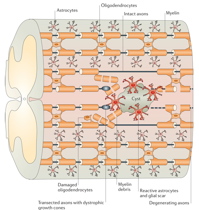
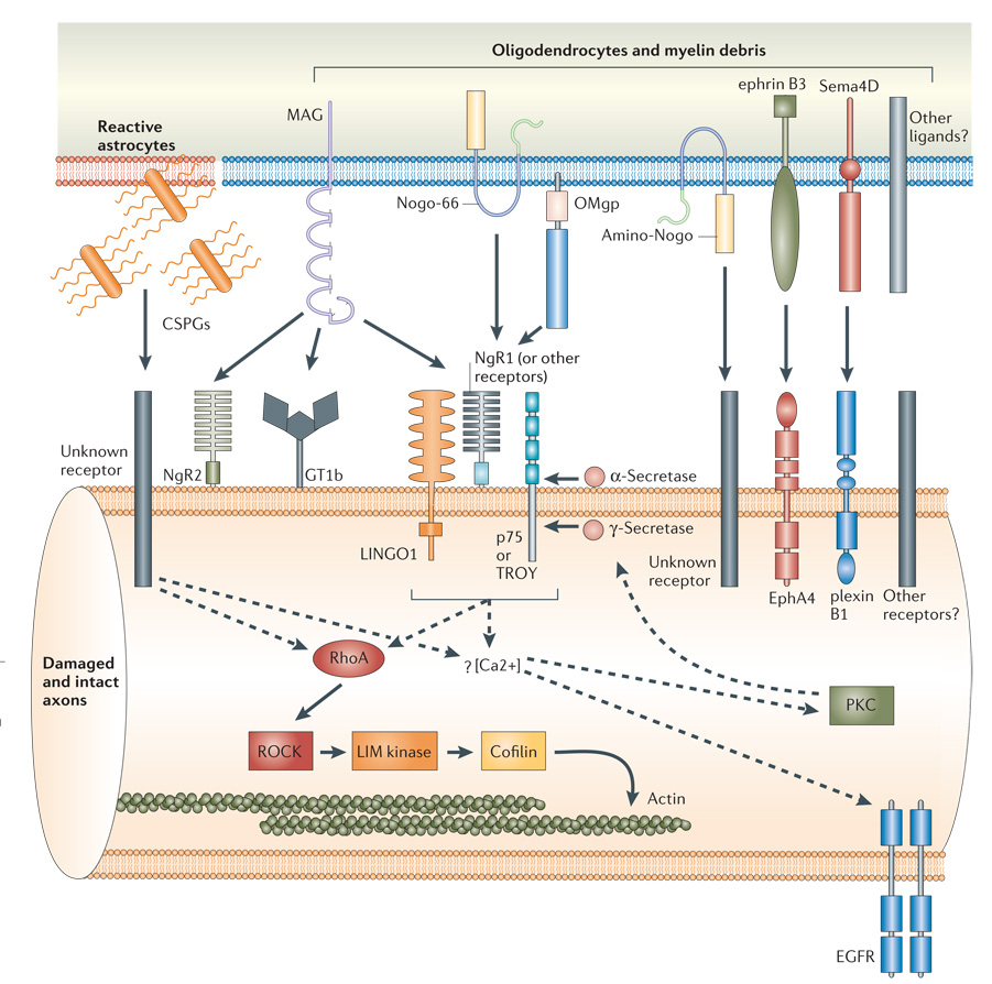
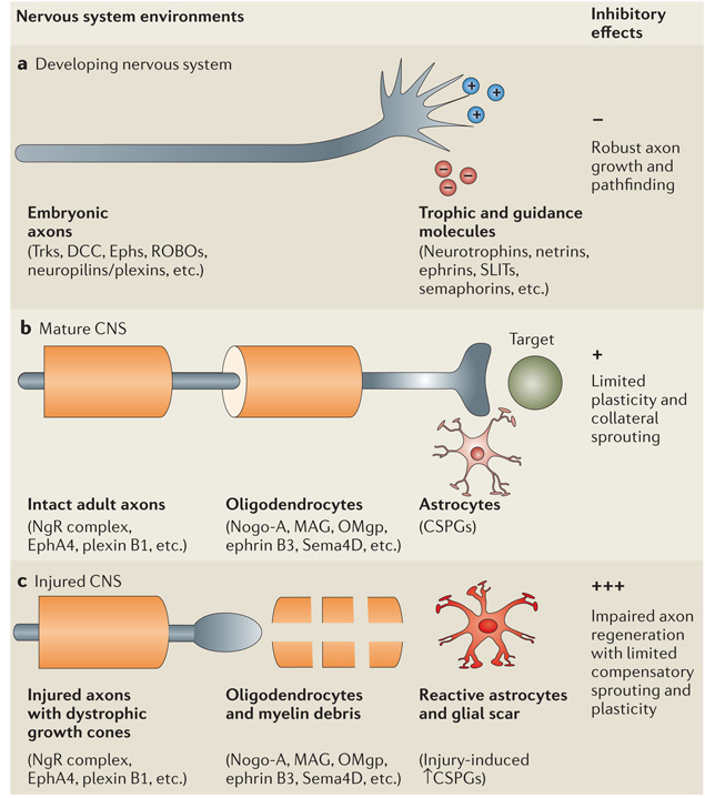
Damage to the adult CNS often leads to persistent deficits due to the inability of mature axons to regenerate after injury. Mounting evidence suggests that the glial environment of the adult CNS, which includes inhibitory molecules in CNS myelin as well as proteoglycans associated with astroglial scarring, might present a major hurdle for successful axon regeneration. Here, we evaluate the molecular basis of these inhibitory influences and their contributions to the limitation of long-distance axon repair and other types of structural plasticity. Greater insight into glial inhibition is crucial for developing therapies to promote functional recovery after neural injury.


Figures

Figure 1
Figure 1. Schematic representation of the CNS injury site
Injury to the adult CNS often results in the transection of nerve fibres and damage to surrounding tissues. The distal ends of the severed axons form characteristic dystrophic growth cones that are exposed to the damaged glial environment. During the early phase of injury, myelin-associated inhibitors from intact oligodendrocytes and myelin debris can restrict axon regrowth–. Recruitment of inflammatory cells and reactive astrocytes over time leads to the formation of a glial scar, often accompanied by a fluid-filled cyst. This scarring process is associated with the increased release of chondroitin sulphate proteoglycans, which can further limit regeneration. Together, these molecular inhibitors of the CNS glial environment present a hostile environment for axon repair.
Figure 2
Figure 2. Glial inhibitors and intracellular signalling mechanisms
The molecular inhibitors of the adult CNS glial environment include chondroitin sulphate proteoglycans (CSPGs) associated with reactive astrocytes from the glial scar, and myelin-associated inhibitors from intact oligodendrocytes and myelin debris, including myelin-associated glycoprotein (MAG),, Nogo-A–, oligodendrocyte myelin glycoprotein (OMgp), ephrin B3 (REF. 26) and the transmembrane semaphorin 4D (Sema4D). Although the topology of Nogo-A remains unclear, both the 66 amino acid loop (Nogo-66) and the amino-terminal domain (amino-Nogo) are known to be inhibitory to axon outgrowth,,,. The neuronal receptors and downstream signalling pathways known to be involved in transducing these inhibitory signals are shown. Among the signalling components that are common to both CSPG and myelin inhibition are the activation of RhoA and the rise in intracellular calcium,,,. Whereas the signals downstream of RhoA that lead to the actin cytoskeleton are well characterized (solid arrows), the relationship between components upstream of RhoA and the role of calcium influx are still ambiguous (dashed arrows). For example, calcium transients might activate protein kinase C (PKC),, which is required for p75 cleavage by γ-secretase, or trigger the transactivation of epidermal growth factor receptor (EGFR).
Figure 3
Figure 3. Changes in CNS environments after maturation and injury
During embryonic development, unmyelinated axons with motile growth cones can extend, retract and respond to various trophic and guidance molecules. This dynamic process allows the neural circuitry to be fine-tuned (a). As the nervous system matures after birth, myelination is finalized with oligodendrocytes ensheathing axons to prevent aberrant sprouting and astrocytes secreting chondroitin sulphate proteoglycans (CSPGs) to further limit structural plasticity in the adult (b). After CNS injury, axons become transected and reactive astrocytes further upregulate their secretion of CSPGs. The distal endings of severed axons form dystrophic growth cones and become exposed to CSPGs from the glial scar, as well as myelin-associated inhibitors from oligodendrocytes and myelin debris (c). As similar mechanisms prevent short-range plasticity in the adult and long-distance axon repair after injury, relieving these inhibitory influences might not only enhance the regeneration of severed axons, but might also promote compensatory sprouting. EphA4, the cognate neuronal receptor for ephrin B3; MAG, myelin-associated glycoprotein; NgR, Nogo-66 receptor; OMgp, oligodendrocyte myelin glycoprotein; Trk, tyrosine receptor kinase.

References

    1. Zito K, Svoboda K. Activity-dependent synaptogenesis in the adult mammalian cortex. Neuron. 2002;35:1015–1017. - PubMed
    1. Zuo Y, Yang G, Kwon E, Gan WB. Long-term sensory deprivation prevents dendritic spine loss in primary somatosensory cortex. Nature. 2005;436:261–265. - PubMed
    1. Edgerton VR, Tillakaratne NJ, Bigbee AJ, de Leon RD, Roy RR. Plasticity of the spinal neural circuitry after injury. Annu. Rev. Neurosci. 2004;27:145–167. - PubMed
    1. Ramón y Cajal S. Degeneration and Regeneration of the Nervous System. London: Oxford Univ. Press; 1928.
    1. Tom VJ, Steinmetz MP, Miller JH, Doller CM, Silver J. Studies on the development and behavior of the dystrophic growth cone, the hallmark of regeneration failure, in an in vitro model of the glial scar and after spinal cord injury. J. Neurosci. 2004;24:6531–6539. An interesting study analysing the morphological changes that constitute the dystrophic growth cones of lesioned axons in the hostile CNS environment.

Publication types