. 2006 Aug 9;25(15):3504-14.
doi: 10.1038/sj.emboj.7601231. Epub 2006 Jul 13.

Involvement of novel autophosphorylation sites in ATM activation

Affiliations

Involvement of novel autophosphorylation sites in ATM activation

Sergei V Kozlov et al. EMBO J. .

Abstract

ATM kinase plays a central role in signaling DNA double-strand breaks to cell cycle checkpoints and to the DNA repair machinery. Although the exact mechanism of ATM activation remains unknown, efficient activation requires the Mre11 complex, autophosphorylation on S1981 and the involvement of protein phosphatases and acetylases. We report here the identification of several additional phosphorylation sites on ATM in response to DNA damage, including autophosphorylation on pS367 and pS1893. ATM autophosphorylates all these sites in vitro in response to DNA damage. Antibodies against phosphoserine 1893 revealed rapid and persistent phosphorylation at this site after in vivo activation of ATM kinase by ionizing radiation, paralleling that observed for S1981 phosphorylation. Phosphorylation was dependent on functional ATM and on the Mre11 complex. All three autophosphorylation sites are physiologically important parts of the DNA damage response, as phosphorylation site mutants (S367A, S1893A and S1981A) were each defective in ATM signaling in vivo and each failed to correct radiosensitivity, genome instability and cell cycle checkpoint defects in ataxia-telangiectasia cells. We conclude that there are at least three functionally important radiation-induced autophosphorylation events in ATM.


Figures
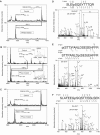
Figure 1
Figure 1
Detection and identification of ATM phosphorylation sites by mass spectrometry. (A) A monoisotopic peptide signal was detected at m/z 1593.8 by MALDI-TOF-MS. This signal was sensitive to treatment with Antarctic phosphatase (PPase). It disappeared, and the signal at m/z 1513.8 (80 U smaller) was stronger after PPase treatment. The signal at m/z 1593.8 matched the theoretical m/z of phospho-ATM363–375 (m/z 1593.7). A meta-stable signal, characteristic of neutral loss of phosphoric acid (−98 U) from a phosphopeptide (shown with an asterisk) confirmed the presence of a phosphopeptide. (B) Signals at m/z 1917.8 and 80 U higher at 1998.0 disappeared after PPase treatment and a new signal at m/z 1837.8 appeared. This shows that the signals at m/z 1917.8 and 1998.0 are mono- and di-phosphorylated forms of the same peptide (80 and 160 U larger, respectively). The insets expand the region from m/z 1996 to 2004 to show that the signal at m/z 1998.0 was removed by PPase treatment. These signals matched the theoretical m/z of mono- and di-phospho-ATM1883–1898 (m/z 1917.8 and 1997.8). (C) A signal at m/z 2080.9 was no longer present after PPase treatment, indicating that it was a phosphopeptide. A signal at 80 U less, at m/z 2000.9, was stronger after PPase treatment. The signal at m/z 2080.9 matched the theoretical mass of phospho-ATM1974–1992 (m/z 2080.9). (D) The phosphopeptide in (A) was sequenced by QqTOF-MSMS. A doubly charged parent ion at m/z 797.4 was selected for fragmentation. The spectrum shows most of the y-type and b-type fragment ions that matched the sequence of ATM363–375 where S367 is phosphorylated (pS). The transition from y8 to y9 demonstrates the presence of a phosphoserine residue. (E) The triply charged parent ion at m/z 639.9 from the phosphopeptide in (B) was selected for fragmentation. The signal intensity in the region from m/z 850 to 1300 has been multiplied by a factor of 8 to improve clarity. The spectrum matches the sequence of phospho-ATM1883–1898. Two independent sequences with different phosphorylation sites were detected. There was sufficient information to confirm that these peptides can be phosphorylated in either of two subdomains: either near its N-terminus (S1883, T1884 or T1885, upper sequence and ions shown with an asterisk) or its C-terminus (S1891 or S1893, lower sequence). The overlapping sequences are demonstrated by the presence of particular fragment ions in both their phosphorylated and non-phosphorylated forms (y11 and y13). (F) The triply charged parent ion of the phosphopeptide in (C) at m/z 694.3 was selected for fragmentation. The spectrum matches the sequence of phospho-ATM1974–1992 where S1981 (pS) is phosphorylated. Multiple y-type and b-type ions rule out the possibility of phosphorylation at any site other than S1981. Note: the parent ions in (D, E) have been truncated in height to improve the clarity of the fragmentation ions.
Figure 2
Figure 2
ATM phosphorylates S367, S1893 and S1981 in vitro. (A) Untreated or irradiated (10 Gy, 1 h) C3ABR lymphoblastoid cells were collected, lysed in the ATM kinase immunoprecipitation buffer (see Materials and methods) and immunoprecipitated with ATM antibody followed by in vitro kinase assays using recombinant GST-ATM 1, 2 and 3 as substrates, containing each of the three tryptic peptides from this figure and Figure 1. (B) Untreated or irradiated (10 Gy, 1 h) C3ABR cells were used in the ATM in vitro kinase assays as described in (A). The following GST substrates were derived from the second tryptic peptide in (B) and Figure 1B (ATM1883–1898) and used in ATM in vitro kinase assays: GST-ATM 4-wild type: ANLDSESEHFFR; GST-ATM 5-S1893A: ANLDSEAEHFFR; and GST-ATM 6-S1891A: ANLDAESEHFFR. (C) Sensitivity of phosphorylation of GST-4 by ATM to wortmannin. Untreated and irradiated (10 Gy, 1 h) C3ABR cells were employed for ATM in vitro kinase activity in the presence or absence of 1 μM wortmannin. (D) A-T (ATIABR) cells are deficient in radiation-induced phosphorylation of GST-4. ATIABR cells contain near-full-length ATM, homozygous for 7636del 9. This is a kinase dead mutant that expresses a less stable form of ATM. In this experiment, equal loading with control was achieved by immunoprecipitating four-fold more cell extracts. Protein kinase activity was determined under the same conditions as described above.
Figure 3
Figure 3
ATM is autophosphorylated on S1893 in response to ionizing radiation. (A) Time course of ATM S1893 and S1981 autophosphorylation after 10 Gy of radiation. Control (C3ABR) lymphoblastoid cells were exposed to 10 Gy of radiation, extracts prepared at indicated time points post-irradiation and immunoprecipitated with anti-ATM antibody before immunoblotting with phosphospecific antibodies against ATM pS1893 and pS1981. A-T represents irradiated AT25ABR. (B) Effect of increasing radiation dose on ATM autophosphorylation on S1893 and S1981. Cells were irradiated and incubated for 60 min before preparation of extracts, immunoprecipitation and immunoblotting as described under panel A. A-T is AT25ABR. (C) Autophosphorylation is not detected in A-T cells with either mutant ATM (ATIABR) or cells not expressing ATM protein (AT25ABR). These cells were irradiated with 10 Gy and incubated for 60 min before processing as described under panel A. C refers to C3ABR cells. Equal loading of ATM mutant was achieved by immunoprecipitating four-fold more protein. (D) Phosphorylation of ATM on S1893 is inhibited by wortmannin. Control (C3ABR) lymphoblastoid cells were pretreated with 20 μM wortmannin for 0.5 h before mock irradiation or exposure to 10 Gy of radiation. Extracts were prepared 1 h post-irradiation and immunoprecipitated with anti-ATM antibody followed by immunoblotting with phosphospecific antibodies against ATM pS1893 and pS1981. (E) Phosphorylation of ATM on S1893 is inhibited by the ATM-specific inhibitor Ku-55933 (KuDOS Pharmaceuticals). C3ABR cells were pretreated with 10 μM Ku-55933 for 1 h before irradiation (10 Gy). Cell extracts were prepared 1 h after irradiation and analyzed as described in panel D.
Figure 4
Figure 4
The Mre11 complex is required for radiation-induced autophosphorylation of ATM in S1893. (A) NBS lymphoblastoid (NBS03LA) and control (C3ABR) cells were exposed to 3 Gy of radiation and extracts prepared for immunoprecipitation and immunoblotting at 15 and 60 min post-irradiation. ATM was immunoprecipitated as in Figure 3A followed by immunoblotting with anti-ATM and anti-pS1981 and pS1893 antibodies. Part of the extract was separated directly in 7.5% SDS–PAGE and immunoblotted for Mre11, Rad50 and Nbs1. As expected, no Nbs1 was detected in NBS03LA cells. (B) A-TLD6 and CBABR lymphoblastoid cells were exposed to 3 Gy of radiation and extracts prepared at 15 and 60 min post-irradiation. ATM was immunoprecipitated followed by immunoblotting with anti-ATM and pS1981 and pS1893 antibodies. A portion of the extracts was directly blotted for Mre11, Rad50 and Nbs1 after separation on SDS–PAGE. ATLD6 is a compound heterozygote with truncating (R571X) and missense (T481K) mutations. The weaker Mre11 band in ATLD6 represents the missense form. Note that both Rad50 and Nbs1 are also expressed at a lower level in those cells, as observed previously (Delia et al, 2004). (C) Rad50-deficient (Ha239) and C3ABR lymphoblastoid cells were exposed to radiation (10 Gy) and incubated for 60 min before preparation of extracts. ATM immunoprecipitated and immunoblotting was carried out as described above. A portion of the extract was also immunoblotted for Mre11, Rad50 and Nbs1. The Ha239 cells contain low levels of Rad50. Again in these cells Mre11 and Nbs1, the other two members of the complex, show reduced levels. Some variability was observed in ATM labeling for both the phospho-S1893 and phospho-S1981 in untreated cells.
Figure 5
Figure 5
Effect of autophosphorylation site mutants on the activation of ATM. (A) Stable cell lines were established in ATIABR cells after transfection with the autophosphorylation site mutants S367A, S1893A and S1981A. Mutant ATM protein expressed from an EBV-based construct (pMEP4) was induced for 18 h with CdCl2 and cells were irradiated with 10 Gy and incubated for a further 10 min. Extracts were prepared and immunoprecipitated with anti-ATM antibody followed by immunoblotting with ATM, pS1893 and pS1981 antibodies. All three mutant proteins were induced to approximately the same amount. The band in the uninduced lanes is owing to endogenous mutant ATM in ATIABR cells. (B) Transfected stable cell lines were induced with CdCl2 before exposure to radiation (10 Gy) and 1 h incubation before preparation of extracts. Full-length ATM (pMAT1) and mutant constructs were employed. In this case, ATM was immunoprecipitated with anti-FLAG antibodies. The pMEP4 ATM constructs all contain a FLAG tag (Zhang et al, 1997). Control (C3ABR) and ATIABR were included for comparison with the transfected cells (right panel). After immunoprecipitation with anti-ATM antibody, immunoblotting was carried out with ATM and downstream substrates p53, pS15-p53, pS343-Nbs1, Nbs1, pT68-Chk2, Chk2, pS957-SMCI and SMCI.
Figure 6
Figure 6
ATM autophosphorylation site mutants are deficient in radiation-induced DNA repair. (A) ATM mutant proteins are defective in formation of radiation-induced phospho-S1981 and γH2AX foci. Stable cell lines expressing full-length ATM (pMAT1) and the three autophosphorylation site mutants were induced with CdCl2 for 18 h, irradiated (10 Gy) and collected after 0.5 and 6 h incubation on slides by cytocentrifugation. Cells were stained with antibodies to phospho-S1981 ATM and γH2AX and analyzed for foci formation using immunofluorescence microscopy. (B) Quantitation of pS1981-ATM and γH2AX foci. The number of both forms of foci per cell was determined in concordance with that observed in the overlays. Merged foci were quantitated and plotted for each cell line at 0.5 and 6 h post-irradiation.
Figure 7
Figure 7
Failure of ATM autophosphorylation site mutants to correct radiosensitivity, genomic instability and cell cycle defects in ATIABR cells. (A) Survival of control (C3ABR), ATIABR and ATIABR cells transfected with pMAT1, S367A, S1893A and S1981A mutant forms of ATM. Cells were induced with CdCl2, exposed to radiation over the range 0–4 Gy and incubated for 3 days, before determination of cell survival. Survival is expressed as a percentage of irradiated/unirradiated. Each point represents an average of triplicate experiments. Error bars represent s.e.m. (B) Radiation-induced (1 Gy) chromosome aberrations in AT1ABR cells transfected with the wild-type and mutant forms of ATM. Cells were induced as described above and aberrations determined as described in Materials and methods. (C) Effect of radiation (1.5 Gy) on the G2/M checkpoint, measured by number of mitotic figures appearing with time after irradiation. Error bars represent s.e.m.

Similar articles

Cited by

References

    1. Ali A, Zhang J, Bao S, Liu I, Otterness D, Dean NM, Abraham RT, Wang XF (2004) Requirement of protein phosphatase 5 in DNA-damage-induced ATM activation. Genes Dev 18: 249–254 - PMC - PubMed
    1. Andegeko Y, Moya L, Mittleman L, Tsarfaty I, Shiloh Y, Rotman G (2001) Nuclear retention of ATM at sites of DNA double strand breaks. J Biol Chem 276: 38224–38230 - PubMed
    1. Bakkenist CJ, Kastan MB (2003) DNA damage activates ATM through intermolecular autophosphorylation and dimer dissociation. Nature 421: 499–506 - PubMed
    1. Beausoleil SA, Jedrychowski M, Schwartz D, Elias JE, Villen J, Li J, Cohn MA, Cantley LC, Gygi SP (2004) Large-scale characterization of HeLa cell nuclear phosphoproteins. Proc Natl Acad Sci USA 101: 12130–12135 - PMC - PubMed
    1. Carney JP, Maser RS, Olivares H, Davis EM, Le Beau M, Yates JR III, Hays L, Morgan WF, Petrini JH (1998) The hMre11/hRad50 protein complex and Nijmegen breakage syndrome: linkage of double-strand break repair to the cellular DNA damage response. Cell 93: 477–486 - PubMed

Publication types

MeSH terms

Substances