. 2006 Jul 26;25(14):3275-85.
doi: 10.1038/sj.emboj.7601230. Epub 2006 Jul 20.

The inner nuclear membrane protein emerin regulates beta-catenin activity by restricting its accumulation in the nucleus

Affiliations

The inner nuclear membrane protein emerin regulates beta-catenin activity by restricting its accumulation in the nucleus

Ewa Markiewicz et al. EMBO J. .

Abstract

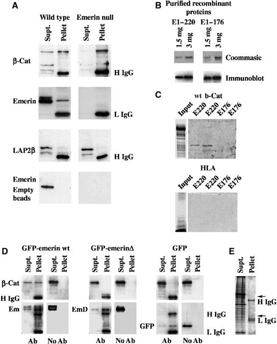
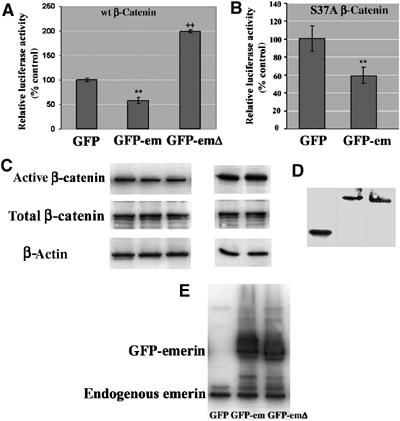
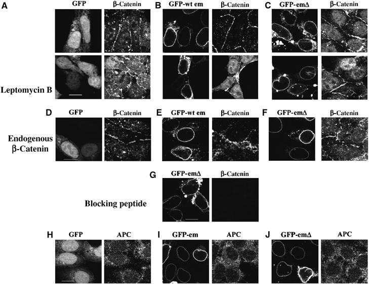
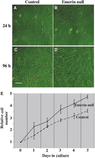
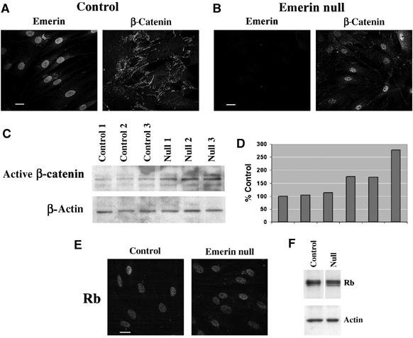
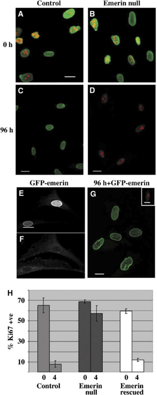
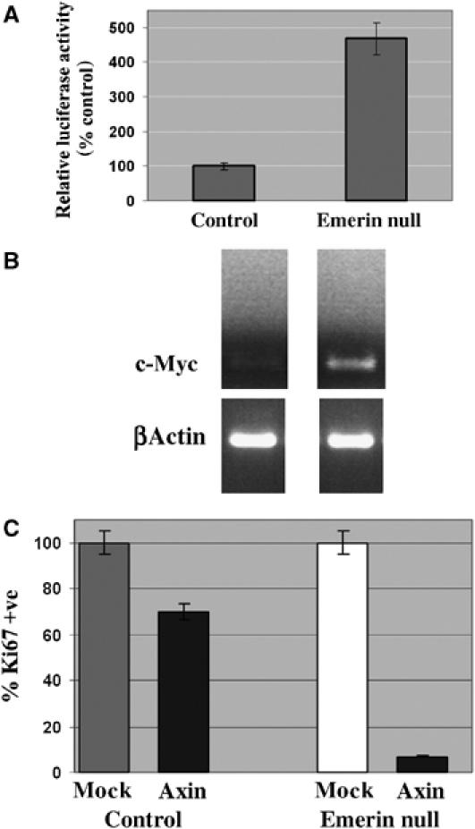
Emerin is a type II inner nuclear membrane (INM) protein of unknown function. Emerin function is likely to be important because, when it is mutated, emerin promotes both skeletal muscle and heart defects. Here we show that one function of Emerin is to regulate the flux of beta-catenin, an important transcription coactivator, into the nucleus. Emerin interacts with beta-catenin through a conserved adenomatous polyposis coli (APC)-like domain. When GFP-emerin is expressed in HEK293 cells, beta-catenin is restricted to the cytoplasm and beta-catenin activity is inhibited. In contrast, expression of an emerin mutant, lacking its APC-like domain (GFP-emerinDelta), dominantly stimulates beta-catenin activity and increases nuclear accumulation of beta-catenin. Human fibroblasts that are null for emerin have an autostimulatory growth phenotype. This unusual growth phenotype arises through enhanced nuclear accumulation and activity of beta-catenin and can be replicated in wild-type fibroblasts by transfection with constitutively active beta-catenin. Our results support recent findings that suggest that INM proteins can influence signalling pathways by restricting access of transcription coactivators to the nucleus.


Figures

Figure 1
Figure 1
β-Catenin binds to emerin through a conserved APC homology domain. (A) β-Catenin was immunoprecipitated from cell extracts prepared from wt or emerin null fibroblasts. Immunoprecipitates were resolved on SDS–PAGE and immunoblotted with antibodies against total β-catenin (β-cat), emerin or LAP2β (top three panels). Alternatively, empty immunobeads were incubated with the same cell extracts and used for immunoblotting with anti-emerin antibody (bottom panel). The antibody used in each blot is indicated to the left-hand side of each panel, while the positions of IgG heavy or light chains (H IgG and L IgG, respectively) are indicated to the right-hand side of each blot. (B) Purified recombinant proteins E1–176 and E1–220 were resolved on SDS–PAGE, which were either stained with Coomassie Brilliant Blue (upper panels) or immunoblotted with anti-emerin antibody (lower panels). (C) 35S-met-labelled wt β-catenin or HLA were resolved on SDS–PAGE and subjected to autoradiography (input). Purified emerin E1–220 and E1–176 at 1.5 and 3 μg were resolved on SDS–PAGE and transferred to nitrocellulose. The filters were then overlayed with 35S-methionine-labelled proteins and subjected to autoradiography. In all panels, the first lane was loaded with 1.5 μg of recombinant protein, while the second lane was loaded with 3.0 μg. (D) β-Catenin and one of GFP-emerin, GFP-emerinΔ or GFP were coexpressed in HEK293 cells. Cell lysates were immunoprecipitated with anti-GFP antibody or empty immunobeads (indicated by Ab or no Ab, respectively, underneath each panel). Immunoprecipitates were immunoblotted with antibodies against β-catenin (upper panels) or GFP (lower panels). The positions of β-catenin (β-cat), GFP-emerin (em), GFP-emerinΔ (emΔ) or GFP are indicated at the left-hand side of each panel. The positions of IgG heavy chain (H IgG) and IgG light chain (L IgG) are indicated at the right-hand side of the final panel. (E) HEK293 cells that were co-transfected with GFP-emerin and β-catenin were immunoprecipitated with anti-GFP antibodies, resolved on SDS–PAGE and stained with ammoniacal silver. In all gels, Supt=material remaining in the supernatant. Pellet=material recovered in immunoprecipitates. Arrow indicates a band with expected mobility of GFP-emerin.
Figure 2
Figure 2
Emerin antagonises β-catenin activity. (A) HEK293 cells were transfected with combinations of wt β-catenin and GFP, GFP-emerin or GFP-emerinΔ together with TOPGLOW or FOPGLOW luciferase reporters and Renilla. (B) Alternatively, HEK293 cells were co-transfected with an activating mutation of β-catenin (S37A) and either GFP or GFP-emerin together with TOPGLOW or FOPGLOW luciferase reporters and Renilla. The levels of luciferase reporter assays were expressed relative to the level of luciferase activity in the presence of GFP. **Significantly reduced luciferase activity (P<0.001), ++Significantly increased luciferase activity (P<0.001). (C) HEK293 cells were transfected with β-catenin and GFP, GFP-emerin or GFP-emerinΔ or S37A and GFP or GFP-emerin, respectively. Cell extracts were prepared for immunoblotting with antibody against active β-catenin, total β-catenin or β-actin. Alternatively, GFP, GFP-emerin or GFP-emerinΔ were expressed in HEK293 cells. Cell lysates were prepared for immunoblotting with either anti-GFP (D) or anti-emerin antibodies (E).
Figure 3
Figure 3
Emerin functions in a β-catenin export pathway. GFP (A), GFP-emerin (B) or GFP-emerinΔ (C) were co-transfected with β-catenin into HEK293 cells. Transfected cultures were grown in the presence or absence of leptomycin B and then fixed and stained with anti-β-catenin antibody followed by TRITC anti-mouse Ig. Alternatively, cultures were transfected with GFP, GFP-emerin and GFP-emerinΔ, then stained with anti-β-catenin antibody to investigate the influence of emerin on endogenous β-catenin (DF). (G) Cells co-transfected with GFP-emerinΔ and β-catenin were stained with anti-β-catenin antibody that had been preabsorbed with a peptide corresponding to its epitope. Finally, cultures transfected with GFP, GFP emerin or GFP-emerinΔ were stained with anti-APC antibody followed by TRITC anti-mouse Ig (HJ). Micrographs show black and white images recorded from each fluorescence channel. Scale bars=10 μm.
Figure 4
Figure 4
Emerin null fibroblasts display a rapid growth phenotype. wt (A, C) or emerin null fibroblasts (B, D) were grown in culture and photographed 24 h (A, B) and 96 h (C, D) after subculture. Alternatively, cells were harvested and counted each day. (E) Shows relative cell numbers after subculture for wt (control) and emerin null fibroblasts over 5 days. Each time point represents pooled data from three different wt and three different emerin null fibroblast strains. At each time point, the relative number for emerin null fibroblasts was always statistically greater than for wt fibroblasts. The P-values were as follows: day 1, 0.03; day 2, 0.05; day 3, 0.001; day 4, 0.005; and day 5, 0.002.
Figure 5
Figure 5
Nuclear accumulation and increased expression of β-catenin in emerin null fibroblasts. Wt (A—control) or emerin null (B) human fibroblasts were costained with antibodies against emerin or active β-catenin to determine the relative distributions of each protein. Micrographs show black and white images taken from each fluorescence channel. Alternatively, extracts were prepared from three different wt and three different emerin null human fibroblast strains and used for immunoblotting with either anti-active β-catenin or anti-β-actin antibodies (C). Densitometry was performed to quantify the level of expression of active β-catenin in each culture relative to the first control culture (D). Wt (control) or emerin null fibroblasts were stained with an antibody against Rb (E) Alternatively, wt (control) or emerin null fibroblasts were prepared for Western blotting using antibodies against total Rb or β-actin (F). Scale bars=10 μm.
Figure 6
Figure 6
Emerin null fibroblasts display autostimulatory growth in culture. Wt or emerin null fibroblasts were transferred to low serum medium. Proliferative capacity was investigated by costaining cultures with either anti-lamin A/C and anti-Ki67 antibodies (A, B) or anti-emerin and anti-Ki67 antibodies (C, D) immediately after transfer to low serum medium (0 h) or 96 h later. Each panel shows two colour merged images in which the green signal represents either lamin A/C or emerin and the red signal represents Ki67. A stable cell line expressing GFP-emerin was established from one emerin null fibroblast strain (EG). The cell line was stained with anti-β-catenin antibody followed by TRITC anti-mouse Ig. The panels show representative black and white images of GFP-emerin (E) and TRITC (F) fluorescence. Alternatively, cultures were transferred to low serum medium for 4 days and then fixed and stained with anti-Ki67 antibody followed by TRITC anti-rabbit Ig (G). The panel shows representative two colour merged images in which GFP-emerin is in green and Ki67 is in red. Inset in (G) shows Ki67 expression in the same cell line before transfer to low serum medium. Scale bars in all micrographs are 10 μm. To quantify the level of expression of Ki67 in each culture used, 200 cells were scored on duplicate slides and in triplicate experiments immediately after transfer to low serum medium (0) and 96 h later (H). The data are expressed as the mean percentage of Ki67 +ve cells±s.e.
Figure 7
Figure 7
β-Catenin activation causes proliferation in emerin null fibroblasts. Wt and emerin null fibroblasts were transfected with β-catenin and either TOPGLOW or FOPGLOW reporters together with Renilla. The levels of luciferase in emerin null fibroblasts are expressed relative to the levels of luciferase in wt fibroblasts (A). RT–PCR was performed on RNA extracted from wt (control) or emerin null fibroblasts using primers specific for c-myc or as a loading control β-actin (B). Wt (control) or emerin null fibroblasts were transfected with FLAG-tagged axin or were mock transfected. After 48 h, cultures were stained with antibodies against Ki67 (or anti-FLAG antibodies to determine transfection efficiency). Two hundred cells were counted on each of triplicate slides to determine the proliferation index. For both control or emerin null cells, the proliferation index in axin-transfected cultures were expressed as a relative percentage of the proliferation index in mock-transfected cultures (C).

References

    1. Behrens J, von Kries JP, Kuhl M, Bruhn L, Wedlich D, Grosschedl R, Birchmeier W (1996) Functional interaction of beta-catenin with the transcription factor LEF-1. Nature 382: 638–642 - PubMed
    1. Bione S, Maestrini E, Rivella S, Mancini M, Regis S, Romeo G, Toniolo D. (1994) Identification of a novel X-linked gene responsible for Emery–Dreifuss muscular dystrophy. Nat Genet 8: 323–327 - PubMed
    1. Bonne G, Di Barletta MR, Varnous S, Becane HM, Hammouda EH, Merlini L, Muntoni F, Greenberg CR, Gary F, Urtizberea JA, Duboc D, Fardeau M, Toniolo D, Schwartz K (1999) Mutations in the gene encoding lamin A/C cause autosomal dominant Emery–Dreifuss muscular dystrophy. Nat Genet 21: 285–288 - PubMed
    1. Brill LM, Salomon AR, Ficarro SB, Mukherjic M, Stettler-Gill M, Peters EC (2004) Robust phosphoproteomic profiling of tyrosine phosphorylation sites from human T cells using immobilised metal affinity chromatography and tandem mass spectrometry. Anal Chem 76: 2763–2772 - PubMed
    1. Cartegni L, di Barletta MR, Barresi R, Squarzoni S, Sabatelli P, Maraldi N, Mora M, Di Blasi C, Cornelio F, Merlini L, Villa A, Cobianchi F, Toniolo D (1997) Heart-specific localization of emerin: new insights into Emery–Dreifuss muscular dystrophy. Hum Mol Genet 13: 2257–2264 - PubMed

Publication types

MeSH terms