. 2006 Jul 26;25(14):3264-74.
doi: 10.1038/sj.emboj.7601228. Epub 2006 Jul 6.

Acetylation and MAPK phosphorylation cooperate to regulate the degradation of active GATA-1

Affiliations

Acetylation and MAPK phosphorylation cooperate to regulate the degradation of active GATA-1

Angel Hernandez-Hernandez et al. EMBO J. .

Abstract

Regulation of transcription requires mechanisms to both activate and terminate transcription factor activity. GATA-1 is a key haemopoietic transcription factor whose activity is increased by acetylation. We show here that acetylated GATA-1 is targeted for degradation via the ubiquitin/proteasome pathway. Acetylation positively signals ubiquitination, suggesting that activation by acetylation simultaneously marks GATA-1 for degradation. Promoter-specific MAPK phosphorylation then cooperates with acetylation to execute protein loss. The requirement for both modifications is novel and suggests a way by which degradation of the active protein can be specifically regulated in response to external phosphorylation-mediated signalling. As many transcription factors are activated by acetylation, we suggest that this might be a general mechanism to control transcription factor activity.


Figures

Figure 1
Figure 1
Mutation of phosphorylation and acetylation sites increases GATA-1 stability. (A) Upper panels: Western blot showing GATA-1 levels in Cos 7 cells, transfected with constructs to express wild-type GATA-1 (Wt), phosphorylation (Ph) or acetylation (Acet) mutants. β-Galactosidase is the cotransfection control. Lower panels: RNase protection assay of the corresponding GATA-1 mRNAs; the human β-globin gene is the cotransfection control. (B) Upper panel: Western blot showing the levels of endogenous and FLAG-tagged GATA-1 protein in BM-SCF cells stably infected with retroviruses to express FLAG-tagged versions of GATA-1. Lower panels: RNase protection assay of the FLAG-tagged and endogenous GATA-1 mRNAs. (C) Pulse–chase experiment to determine the half-life of GATA-1. The levels of wild type (⧫), acetylation (Acet ▪) and phosphorylation (Ph ▴) mutants in Cos 7 cells are shown. A value of 100% was given to the amount of protein at time zero; the per cent of protein remaining was calculated relative to this. The plot shows the average of three experiments; error bars show standard error.
Figure 2
Figure 2
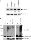
GATA-1 is ubiquitinated. (A) Proteasome inhibitors increase GATA-1 stability. Transfected Cos 7 cells were treated with the proteasome inhibitors shown. Protein levels were analysed by immunoblotting with N6 antibody; β-galactosidase is the cotransfection control. (B) Phosphorylation and acetylation affect GATA-1 ubiquitination. Western blot showing the level of ubiquitination of GATA-1 immunoprecipitated from transfected 293T cells. Normalised levels of GATA-1 were loaded; this is verified in the lower panel where the blot is re-probed with a second anti-GATA-1 antibody; densitometry confirmed GATA-1 ratios as wt 1:Ph mut 0.8:Acet mut 0.95. Lanes 5 and 6 are a short exposure to highlight the difference in GATA-1 ubiquitination ±MG-132. Mock-transfected cells confirm that ubiquitinated proteins do not stick nonspecifically during immunoprecipitation.
Figure 3
Figure 3
Acetylation of GATA-1 causes ubiquitination. (A) Treatment with TSA increases acetylated and ubiquitinated GATA-1. Upper panel: Western blot showing GATA-1 levels in whole-cell extract from Cos 7 cells that had been transfected with GATA-1 144D (Supplementary Figure 1B) and β-galactosidase with (+) or without (−) TSA treatment. Lower panel: Western blot showing the levels of ubiquitinated GATA-1 (Ub-GATA-1), acetylated GATA-1 (Ac-GATA-1) and total GATA-1 following immunoprecipitation from transfected Cos 7 cells treated with MG-132 with (+) or without (−) TSA. (B) p300 but not p300dHAT decreases GATA-1 levels. NIH3T3 cells were transfected with expression vectors for GATA-1 and p300 or its acetyltransferase mutant, p300dHAT. Whole-cell extracts were used in a Western blot with anti-GATA-1 antibody. β-Galactosidase is the cotransfection control. (C) The decrease in GATA-1 levels upon TSA treatment in haemopoietic (BM-SCF) cells is reversed by MG-132. As for part A except that endogenous GATA-1 was examined. Upper panel: Detection of acetylated GATA-1; second panel: detection of total GATA-1. The right panel shows ubiquitinated and total GATA-1 in the presence of MG-132. Phosphorylated total GATA-1 is indicated by P-GATA-1 and phosphorylated, acetylated GATA-1 by P/Ac-GATA-1. Lambda phosphatase confirmed that the slower mobility form is phosphorylated GATA-1 (Supplementary Figure 2E). Total GATA-1 levels decrease more in Cos 7 cells (A, upper panel) than BM-SCF cells (C, second panel) upon TSA treatment. GATA-1 regulates its own promoter in haemopoietic cells but is controlled by the EF1α promoter in Cos 7 cells. To prevent TSA increasing GATA-1 levels in BM-SCF cells via GATA-1 autoregulation, we added the transcription inhibitor α-amanitin (α-aman). A more substantial decrease is now seen (fourth panel). An anti-β-tubulin Western confirms that equivalent cell numbers were used (lower panel). (D) All acetylation mutants are more stable than the wild-type protein. Groups of acetylation sites were mutated (K to R). N: K245, K246 and K252; C: K308, K312 and K316; N+C1: K245, K246, K252, K312, K314 and K316; N+C2: all N and C mutations. Acet mut is that used in other experiments. The level of GATA-1 protein was determined by Western blotting of whole-cell extracts from transfected 293T cells. β-Galactosidase is the cotransfection control. Mutation of two different sets of three lysines (that are substrates for acetylation; Boyes et al, 1998) resulted in a similar increase in GATA-1 stability, suggesting that it is unlikely that loss of ubiquitination sites caused the increased stability. (E) The phosphorylation mutants are acetylated. GATA-1 phosphorylation mutant proteins (serine to alanine) were immunoprecipitated from transfected 293T cells; a Western blot probed with anti-acetyl-GATA-1 antibody is shown. Equivalent amounts of immunoprecipitated GATA-1 were loaded; this was confirmed by re-probing with a second anti-GATA-1 antibody (lower panel). The anti-acetyl-GATA-1 antibody was raised against amino acids 307–317; the lower signal for S310A and total phosphorylation mutant is probably because some antibodies in the polyclonal mix require S310 to be wild type. A lower signal was not seen with an antibody raised against all acetylated lysines (Supplementary Figure 2C).
Figure 4
Figure 4
Phosphorylation and acetylation cooperate to cause the degradation of acetylated GATA-1. (A) Each individual phosphorylation mutant is more stable than wild-type protein. Western blot showing GATA-1 phosphorylation mutant proteins immunoprecipitated from transfected Cos 7 cells. Transfection efficiency was normalised to β-galactosidase. (B) Each phosphorylation mutant is less ubiquitinated than wild-type protein. The levels of ubiquitination were determined via immunoprecipitation from transfected 293T cells as described in Figure 2. The same Western blot was re-probed with anti-GATA-1 antibody (lower panel) and confirms that protein is present in all lanes. Higher ubiquitination of the total phosphorylation mutant and S26A (which is similar between the two proteins but notably less than wild type) correlates with their lower protein levels (Figure 4A). (C) Phosphorylation is needed for efficient degradation of acetylated GATA-1. Western blot showing GATA-1 levels in extracts from BM-SCF cells that stably express GATA-1 mutated in serine 26 (Flag-26A). Endogenous GATA-1 in the same cell provides an internal control for degradation upon TSA treatment. β-Tubulin levels verify that equivalent cell numbers were taken (lower panel). (D) MEK inhibitor prevents degradation of acetylated GATA-1. Haemopoietic (BM-SCF) cells were treated with or without TSA in the presence or absence of the MEK inhibitor U0126. Acetylated GATA-1 (Ac-GATA-1; upper panel) and total GATA-1 (GATA-1; lower panel) were detected as in Figure 3. The graph shows quantification of the level of acetylated GATA-1 divided by the amount of total GATA-1 in two further independent experiments where a higher level of acetylated GATA-1 in untreated cells was observed.
Figure 5
Figure 5
Preferential degradation of transcriptionally active GATA-1. (A) Upper: The reporter construct pGL3EpoR. Lower: Inhibition of phosphorylation increases GATA-1-dependent transcription. NIH3T3 cells, transfected with the constructs shown, were treated with or without the MEK inhibitor U0126. The transcription level in the presence of U0126 divided by that in its absence is plotted. The average of four experiments is shown. (B) The phosphorylation mutant is more transcriptionally active than wild-type GATA-1. Left panel: S1 mapping of β-globin gene transcription in BM-SCF cells that express FLAG-tagged wild-type (wt) or phosphorylation mutant (ph mut) GATA-1. Quantification of β-globin transcription, normalised to α-actin, is shown beneath the gel. Right panel: Western blot showing the level of the FLAG-tagged wild-type or phosphorylation mutant protein and endogenous GATA-1. For this experiment, cell lines were chosen where mutant and wild-type GATA-1 are expressed at similar levels. (C) The RNRK mutant does not bind DNA. Equivalent amounts of whole-cell extract prepared from mock-transfected Cos 7 cells (C) or cells that had been transfected with expression vectors for wild-type (wt) or mutant GATA-1 proteins (IQT and RNRK) were used in a gel mobility shift assay under the conditions described (Boyes et al, 1998). Control experiments confirmed that transfection efficiency was equivalent. (D) The DNA binding mutants are more stable than wild-type GATA-1. Left panel: Western blot of extracts from Cos 7 cells transfected with wild-type GATA-1 and the DNA binding mutants, C261P and RNRK, probed with anti-GATA-1 antibody or anti-acetyl-GATA-1 antibody. β-Galactosidase is the cotransfection control. Right panel: Haemopoietic (BM-SCF) cells, expressing FLAG-tagged RNRK GATA-1, were treated with or without TSA. A Western blot probed with anti-GATA-1 antibody (N-6) is shown.
Figure 6
Figure 6
DNA binding is required for MAPK phosphorylation but not acetylation of GATA-1. (A) DNA binding inhibits acetylation of GATA-1. A purified peptide of the double zinc-finger region of GATA-1 was either bound or unbound to an oligonucleotide with a palindromic GATA-1 binding site and an in vitro acetylation reaction with p300 was performed (Boyes et al, 1998). At this binding site, the half-life of GATA-1/DNA binding is 70 min and thus should persist during the acetylation reaction. The stained gel (left) shows that equivalent amounts of protein were loaded. The autoradiograph (right) shows acetylation of GATA-1 and p300. (B) DNA binding is required for phosphorylation of GATA-1 at S26 and S178. Phosphopeptide maps of wild-type GATA-1 and RNRK mutant protein that had been immunoprecipitated from transfected, 32P-labelled Cos 7 cells and then digested with chymotrypsin or trypsin as indicated are shown. The sites of phosphorylation were identified from Crossley and Orkin (1994). One spot showing phosphorylation at S142 is smeared in the mutant protein; as the other spot is present at normal intensity, we suggest that phosphorylation at S142 is unaffected by DNA binding. The spot previously correlated with phosphorylation at S310 is visible in the trypsin digests. For the DNA binding mutant, this spot is shifted slightly to the left, indicative of a higher molecular weight. We suggest that this alteration in tryptic digestion pattern is owing to loss of a cleavage site in the RNRK mutant. (C) The RNRK mutant protein is localised to the nucleus. Nuclear and cytoplasmic extracts were prepared (Supplementary methods) from BM-SCF cells, which stably express FLAG-tagged RNRK mutant. An equivalent volume of each extract was analysed by Western blotting using anti-GATA-1 and anti-β-tubulin antibodies. (D) The RNRK mutant protein can be phosphorylated. Cos 7 cells were transfected with expression vectors for the GATA-1 proteins shown either with or without an expression vector for the constitutively active MAP kinase (CA-MAPKK). Whole-cell extract was used in a Western blot with anti-GATA-1 antibody (N6). Upper panel: The amount of extract used was varied to achieve more equivalent GATA-1 levels for ease of comparison of the phosphorylation shift. Lower panel: An equivalent volume of extract from each transfection was used and indicates that expression of CA-MAPKK causes loss of GATA-1. (E) GATA-1 binding to the EKLF enhancer is increased when MAPK signalling is inhibited. Chromatin immunoprecipitation was performed in BM-SCF cells following treatment with (UO126) or without (control) MEK inhibitor. The DNA was analysed by real-time PCR using primers spanning a GATA-1 site in the EKLF enhancer (positive site) and a negative control site. The fold enrichment compared to input Ct values is shown.
Figure 7
Figure 7
Regulation of GATA-1 ubiquitination by SCF and erythropoietin. (A) GATA-1 ubiquitination is increased in response to signalling by SCF and erythropoietin. BM-SCF or BM-Epo cells were starved of cytokine for 6 h. SCF or erythropoietin was then re-administered and GATA-1 immunoprecipitated at the times shown. GATA-1 ubiquitination was analysed with an anti-ubiquitin antibody and GATA-1 levels with an anti-GATA-1 antibody (M-20; middle panel). Sixty minutes after induction, GATA-1 levels increased, most likely owing to new GATA-1 synthesis. Phosphorylated GATA-1 (P-GATA-1) is indicated. LT indicates cells grown continuously (long term) in SCF. β-Tubulin levels verify that equivalent cell numbers were used (lower panel). (B) Phosphorylation is required for SCF-induced GATA-1 turnover. BM-SCF cells that express FLAG-tagged GATA-1 mutated in individual phosphorylation sites (S26A, S49A and S72A) were induced with SCF. Immunoblotting with anti-FLAG antibody showed that SCF did not cause a phosphorylation shift or loss of GATA-1. FLAG-tagged wild-type GATA-1 is lost like the endogenous protein (compare lower panel and panel A middle panel). (C) Inhibition of phosphorylation prevents GATA-1 ubiquitination. BM-SCF cells were starved of growth factor for 6 h. SCF was then re-administered either in the absence (SCF) or presence (SCF+U0126) of the MEK inhibitor U0126. Samples were taken at the times indicated, immunoprecipitated with anti-GATA-1 antibody and the level of ubiquitination determined via Western blotting using an anti-ubiquitin antibody. The middle panel shows GATA-1 levels following re-probing of the blot with a second anti-GATA-1 antibody (M-20). Phosphorylated and non-phosphorylated GATA-1 are indicated by asterisks. (D) Acetylation is required for GATA-1 turnover. Upper panel: GATA-1 acetylation mutant is not degraded in response to SCF signalling; endogenous GATA-1 in the same cell is lost (second panel from top). Fourth panel: Acetylated GATA-1 (Ac-GATA-1) is degraded faster than bulk GATA-1 (lower panel, where the same blot was re-probed) upon mitogen signalling.

Similar articles

Cited by

References

    1. Blobel GA, Nakajima T, Eckner R, Montminy M, Orkin SH (1998) CREB-binding protein cooperates with transcription factor GATA-1 and is required for erythroid differentiation. Proc Natl Acad Sci USA 95: 2061–2066 - PMC - PubMed
    1. Boyes J, Bird A (1992) Repression of gene by DNA methylation depends on CpG density and promoter strength:evidence for involvement of a methyl-CpG binding protein. EMBO J 11: 327–333 - PMC - PubMed
    1. Boyes J, Byfield P, Nakatani Y, Ogryzko V (1998) Regulation of activity of the transcription factor GATA-1 by acetylation. Nature 396: 594–598 - PubMed
    1. Cairns L, Ciro M, Minuzzo M, Morle F, Starck J, Ottolenghi S, Ronchi A (2003) Induction of globin mRNA expression by interleukin-3 in a stem cell factor-dependent SV-40 T-antigen-immortalized multipotent hematopoietic cell line. J Cell Physiol 195: 38–49 - PubMed
    1. Chi Y, Huddleston MJ, Zhang X, Young RA, Annan RS, Carr SA, Deshaies RJ (2001) Negative regulation of Gcn4 and Msn2 transcription factors by Srb10 cyclin-dependent kinase. Genes Dev 15: 1078–1092 - PMC - PubMed

Publication types

Substances