Comparative Study
. 2006 Jul 26;25(14):3389-97.
doi: 10.1038/sj.emboj.7601219. Epub 2006 Jul 6.

Human ABH3 structure and key residues for oxidative demethylation to reverse DNA/RNA damage

Affiliations
Comparative Study

Human ABH3 structure and key residues for oxidative demethylation to reverse DNA/RNA damage

Ottar Sundheim et al. EMBO J. .

Abstract

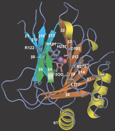
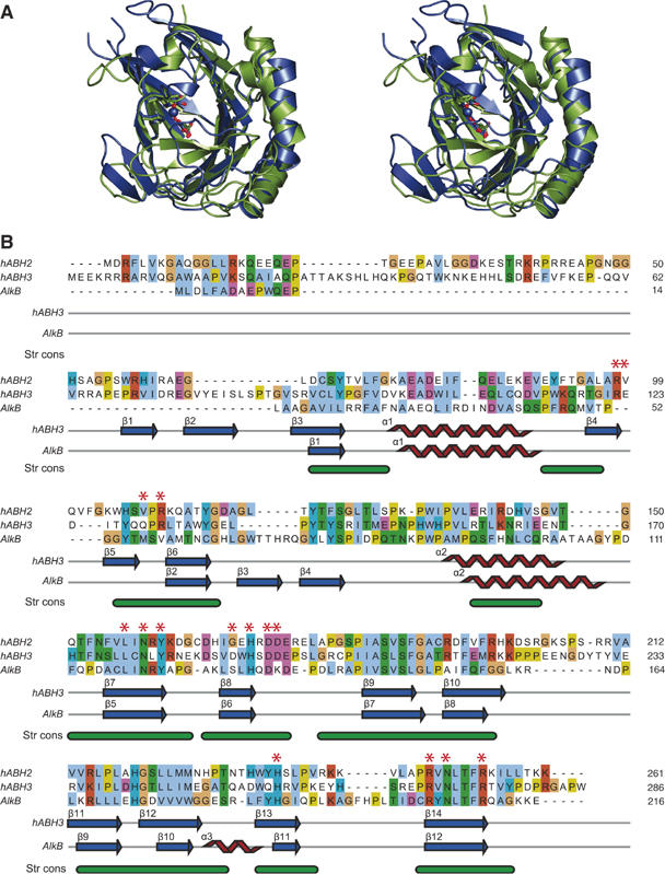
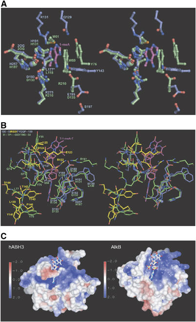
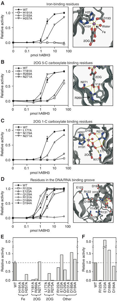
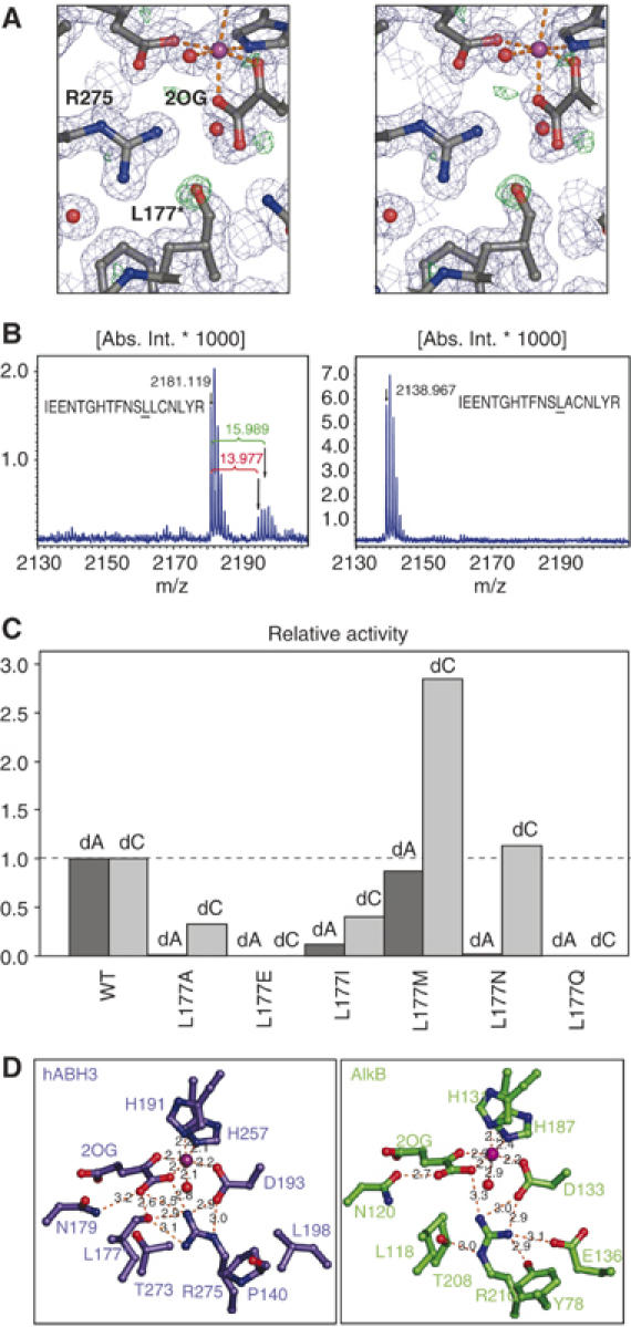
Methylating agents are ubiquitous in the environment, and central in cancer therapy. The 1-methyladenine and 3-methylcytosine lesions in DNA/RNA contribute to the cytotoxicity of such agents. These lesions are directly reversed by ABH3 (hABH3) in humans and AlkB in Escherichia coli. Here, we report the structure of the hABH3 catalytic core in complex with iron and 2-oxoglutarate (2OG) at 1.5 A resolution and analyse key site-directed mutants. The hABH3 structure reveals the beta-strand jelly-roll fold that coordinates a catalytically active iron centre by a conserved His1-X-Asp/Glu-X(n)-His2 motif. This experimentally establishes hABH3 as a structural member of the Fe(II)/2OG-dependent dioxygenase superfamily, which couples substrate oxidation to conversion of 2OG into succinate and CO2. A positively charged DNA/RNA binding groove indicates a distinct nucleic acid binding conformation different from that predicted in the AlkB structure with three nucleotides. These results uncover previously unassigned key catalytic residues, identify a flexible hairpin involved in nucleotide flipping and ss/ds-DNA discrimination, and reveal self-hydroxylation of an active site leucine that may protect against uncoupled generation of dangerous oxygen radicals.


Figures

Figure 1
Figure 1
hABH3 overall structure and active site location. Ribbon representation of hABH3Δ69 with secondary structure assignments. The β-strand jelly roll is made up by two antiparallel β-sheets coloured orange and blue, respectively. The β4–β5 hairpin is coloured green. Helices are coloured yellow, and key residues as well as the co-substrate 2OG are shown as balls and sticks. The iron (violet) and the iron bound water (red) are shown as spheres.
Figure 2
Figure 2
The overall hABH3 β-strand jelly roll core resembles AlkB. (A) Stereo pair ribbon representation of hABH3Δ69 (blue) superimposed upon AlkB (2FDJ) (green). The side chains in the His1-X-Asp-Xn-His2 motif and the 2OG (hABH3) and succinate (AlkB) are shown as balls and sticks, whereas the iron ions are presented as spheres. (B) Sequence alignment and secondary structure assignment of AlkB, hABH2 and hABH3. The alignment of hABH3 and AlkB was generated with DeepView (Guex and Peitsch, 1997) based on a structural alignment of the 3D structures. The hABH2 sequence was added to the alignment by a combination of profile alignment of hABH2 versus the structure-based hABH3/AlkB alignment, using ClustalX (Thompson et al, 1997), as well as hABH2/hABH3 and hABH2/AlkB alignments from iterative PSI-Blast searches (Altschul et al, 1997) starting from hABH2. The final alignment was made with JalView (Clamp et al, 2004). The bottom annotation line (Str cons) shows structurally conserved regions (in green) identified by DeepView. Individual residues are coloured according to ClustalX colour coding, and residues selected for site-directed mutation analysis are indicated by red asterisks.
Figure 3
Figure 3
Key regions in hABH3 and their differences from AlkB. (A) Stereo pair ball and stick representation of active site residues in hABH3 (blue) and AlkB (green). The 1-meA lesion from the AlkB complex is coloured magenta. The substrate-binding pocket of hABH3 is dominated by polar residues, while the observed substrate interactions in AlkB are predominantly hydrophobic. The oxidised Leu177 is denoted L177*. (B) Stereo pair representation of the active site entrances of hABH3 (blue) and AlkB (green). The structurally nonconserved residues in the β4–β5 loop (upper left) and the β6–α2 region (lower left) of hABH3 are in yellow. (C) Electrostatic surface presentations of hABH3 and AlkB contoured at ±2 kT/e, where red and blue describe negative and positive potentials, respectively. Electrostatic potentials were calculated by UHBD (Gilson et al, 1993), and are mapped on the molecular surface using AVS (Advanced Visual Systems Inc.). The T-1-meA-T (balls and sticks) was positioned in hABH3 by superimposition of the AlkB/trinucleotide complex upon hABH3.
Figure 4
Figure 4
Active site residue structure–function relationships and enzyme activity shown by WT and mutant hABH3-mediated release of radioactivity from [3H]methyl DNA. Left: (AD) Relative 1-meA repair activity for full-length WT hABH3 and alanine mutants thereof at 0.1, 0.3, 1, 3, 10 and 50 pmol enzyme. Mutants are grouped according to the functions of different residues. Assay reproducibility was verified by including the WT in each experiment comprising three mutants, so every activity curve data point for WT is based on four independent experiments each in duplicate, with standard deviation indicated. Each point in the graphs for mutants represents the mean of duplicates. Right: (A–D) Closeup presentations of the residues in hABH3 analysed by site-directed mutation. The iron ion (violet) and iron-bound water (red) are shown as spheres. (E) Activity of hABH3 mutants involved in iron-binding, 2OG C-5 carboxylate binding, 2OG C-1 carboxylate binding, and residues in the DNA/RNA binding cleft relative to WT on 1-meA-containing ssDNA using 0.3, 1, 3 and 10 pmol enzyme. Bars represent average activity from the four enzyme concentrations relative to WT. (F) Average activity of WT and mutants Arg122Ala, Glu123Ala, Asp189Ala and Asp194Ala, acting upon 1-meA-containing dsDNA substrate using 0.3, 1, 3 and 10 pmol enzyme.
Figure 5
Figure 5
Analysis of Leu177 oxidation and activity of Leu177 mutants. (A) Stereo view of the electron density around the hABH3 active site residues, 2OG and iron. 2FoFc (contoured at 1σ, in blue) and FoFc (contoured at 3σ, in green) were calculated after omitting the Leu177 modification (assigned Oɛ). In the crystal structure, the modification was modelled as carbonyl (L177*) with a refined distance of 1.23 Å between the Leu177 Cδ2 and Oɛ refined to a Cγ-Cδ2-Oɛ angle of 119.6°. (B) The modification gave mass-shifts of +14 and +16 Da in MALDI-TOF MS, revealing a mixture of carbonyl and hydroxyl at Cδ2. (C) Relative activity of Leu177 mutants using [3H]methylated poly(dA) and poly(dC) ssDNA substrates. Activity assays were performed in duplicate using 5 pmol of each hABH3 mutant. Data for Leu177Ala are the same as presented in Figure 4E. (D) Closeup view of the 2OG-coordinations in hABH3 and AlkB. The essential Arg275 in hABH3 adapts a different conformation than the corresponding Arg210 in AlkB, and makes a hydrogen bond to the oxidised Leu177.

References

    1. Aas PA, Otterlei M, Falnes PO, Vagbo CB, Skorpen F, Akbari M, Sundheim O, Bjoras M, Slupphaug G, Seeberg E, Krokan HE (2003) Human and bacterial oxidative demethylases repair alkylation damage in both RNA and DNA. Nature 421: 859–863 - PubMed
    1. Altschul SF, Madden TL, Schaffer AA, Zhang J, Zhang Z, Miller W, Lipman DJ (1997) Gapped BLAST and PSI-BLAST: a new generation of protein database search programs. Nucleic Acids Res 25: 3389–3402 - PMC - PubMed
    1. Aravind L, Koonin EV (2001) The DNA-repair protein AlkB, EGL-9, and leprecan define new families of 2-oxoglutarate- and iron-dependent dioxygenases. Genome Biol 2: 7.1–7.8 - PMC - PubMed
    1. Barrett TE, Savva R, Panayotou G, Barlow T, Brown T, Jiricny J, Pearl LH (1998) Crystal structure of a G:T/U mismatch-specific DNA glycosylase: mismatch recognition by complementary-strand interactions. Cell 92: 117–129 - PubMed
    1. Bjelland S, Bjoras M, Seeberg E (1993) Excision of 3-methylguanine from alkylated DNA by 3-methyladenine DNA glycosylase I of Escherichia coli. Nucleic Acids Res 21: 2045–2049 - PMC - PubMed

Publication types

MeSH terms

Associated data