. 2006 Jul 26;25(14):3245-56.
doi: 10.1038/sj.emboj.7601214. Epub 2006 Jul 6.

Dynamic molecular confinement in the plasma membrane by microdomains and the cytoskeleton meshwork

Affiliations

Dynamic molecular confinement in the plasma membrane by microdomains and the cytoskeleton meshwork

Pierre-François Lenne et al. EMBO J. .

Abstract

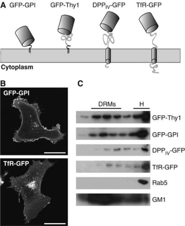
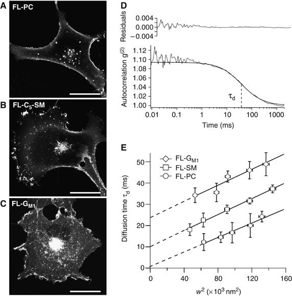
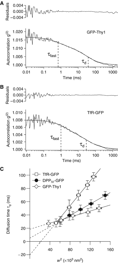
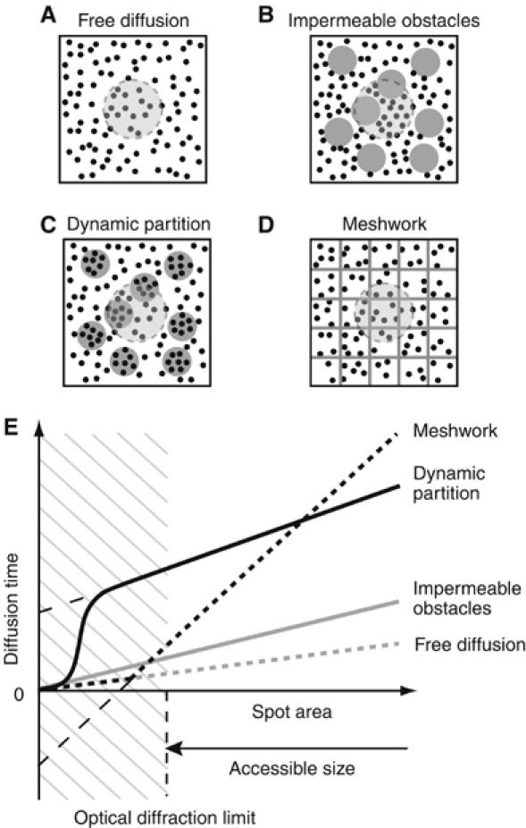
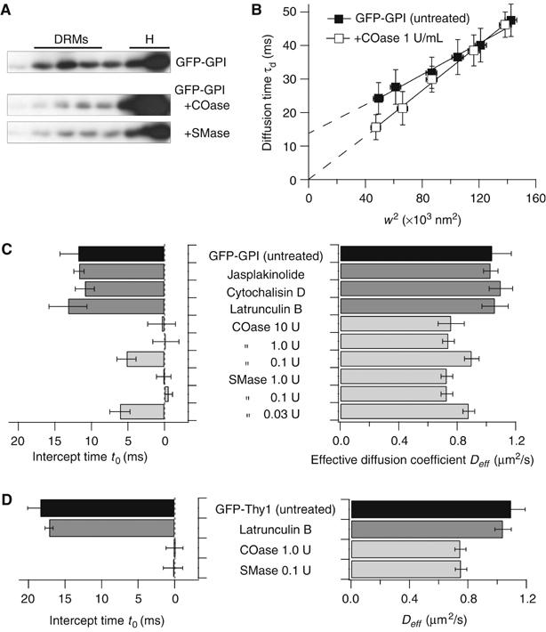
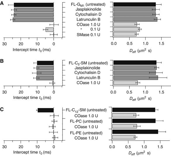
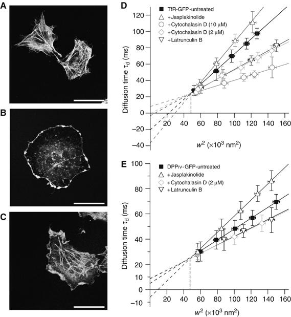
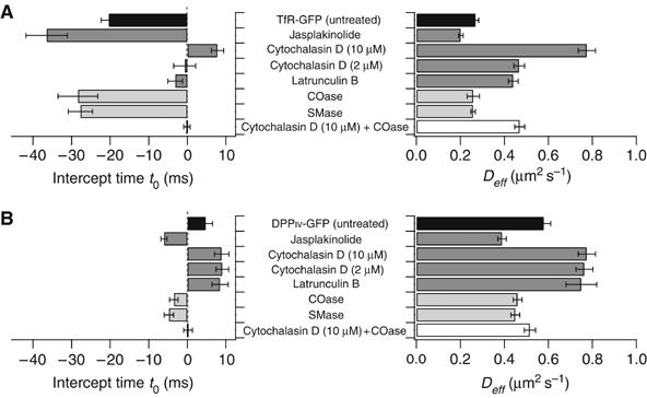
It is by now widely recognized that cell membranes show complex patterns of lateral organization. Two mechanisms involving either a lipid-dependent (microdomain model) or cytoskeleton-based (meshwork model) process are thought to be responsible for these plasma membrane organizations. In the present study, fluorescence correlation spectroscopy measurements on various spatial scales were performed in order to directly identify and characterize these two processes in live cells with a high temporal resolution, without any loss of spatial information. Putative raft markers were found to be dynamically compartmented within tens of milliseconds into small microdomains (Ø <120 nm) that are sensitive to the cholesterol and sphingomyelin levels, whereas actin-based cytoskeleton barriers are responsible for the confinement of the transferrin receptor protein. A free-like diffusion was observed when both the lipid-dependent and cytoskeleton-based organizations were disrupted, which suggests that these are two main compartmentalizing forces at work in the plasma membrane.


Figures

Figure 1
Figure 1
GFP-tagged proteins used in this study. (A) Membrane topology of the GFP-tagged proteins. (B) Confocal images of COS-7 cells expressing the GFP-GPI and TfR-GFP proteins. Scale bar is 20 μm. (C) Brij 98 solubilized PNS from transfected COS-7 cells was fractionated on the sucrose gradient and blotted with anti-GFP, anti-Rab 5 antibodies or cholera toxin B.
Figure 2
Figure 2
Diffusion behavior of lipid analogs. Confocal images of COS-7 cells after labeling with lipid analogs: (A) FL-PC, (B) FL-SM and (C) FL-GM1. Scale bar is 20 μm. (D) ACF obtained with FL-GM1 lipids. The curve was satisfactorily fitted with a 1-species free diffusion model (see Supplementary data). The diffusion time τd was given by the lag time at half maximum. (E) Diffusion behavior of lipid analogs. Confinement times were determined from the position at which the diffusion curves and the time-axis intersected. Error bars in x and y give standard deviations (s.d.) of the means.
Figure 3
Figure 3
Diffusion behavior of GFP-tagged proteins. ACFs obtained with GFP-Thy1 (A) and TfR-GFP (B). In both cases, two decay times were clearly identified with the various probes used: a short time τfast and a longer one τd. (C) FCS diffusion laws in the case of TfR-GFP, DPPIV-GFP and GFP-Thy1. Error bars on the x and y axes give the s.d.s of the means.
Figure 4
Figure 4
Simulated FCS diffusion laws corresponding to different membrane models. (A–D) Diffusion models for membrane organization. (A) In the free diffusion model, fluorescent molecules (black dots) show pure Brownian motion and fluoresce under the laser excitation spot (large gray circle). (B) In the presence of impermeable obstacles (gray spots), the diffusion is restricted to the free space. (C) When the domains are permeable (as the result of dynamic partition processes), the molecules can diffuse into and out of the microdomains and be transiently trapped (gray spots). (D) In a meshwork model, multiple adjacent domains are separated by barriers (gray lines) preventing the diffusion of the molecules. (E) Characteristic diffusion laws obtained for the different models. The diffusion time τd was analyzed as a function of the squared radius w2. Diffusion laws applying to the free diffusion model (dotted gray curve), the diffusion process in the presence of impermeable obstacles (gray curve), the dynamic partition model (dotted black curve) and the meshwork model (black curve). Diffusion laws cannot be determined experimentally below the diffraction limit. For detailed analyses, see the Supplementary data and Wawrezinieck et al (2004, 2005).
Figure 5
Figure 5
The confinement of GFP-tagged GPI-anchored proteins in microdomains is lipid-dependent. (A) Biochemical determination of GFP-GPI localization in DRMs after COase or SMase treatment. (B) FCS diffusion laws governing GFP-GPI proteins after cell treatment with COase. Error bars on the x and y axes are the s.d.s of the means. (C) Time intercept t0 and effective diffusion coefficient Deff obtained with GFP-GPI in untreated COS-7 cells (black bars), after cytoskeleton drug treatments (dark gray bars) or lipid modifications (light gray bars). (D) Idem in the case of GFP-Thy1 protein.
Figure 6
Figure 6
The confinement of lipid probes in the plasma membrane of COS-7 cells is lipid-dependent. Time intercept t0 and effective diffusion coefficient Deff obtained with FL-GM1 (A), FL-C5-SM (B) and the other lipid analogs used in this study (C) in untreated COS-7 cells (black bars), after applying cytoskeleton drug treatment (dark gray bars) or lipid modifications (light gray bars). Error bars give the s.d.s of the means.
Figure 7
Figure 7
The GFP-tagged transmembrane protein diffusion laws are actin-dependent. Confocal images of rhodamine phalloidin labeling of F-actin in COS-7: (A) untreated; (B) 1 μM latrunculin B; (C) 0.4 μM jasplakinolide. Diffusion laws governing TfR-GFP (D) and DPPIV-GFP (E) after applying cytoskeleton drug treatment. Error bars on the x and y axes are the s.d.s of the means.
Figure 8
Figure 8
Both actin meshwork and lipid domains contribute to transmembrane protein compartmentalization. Time intercept t0 and effective diffusion coefficient Deff obtained with TfR-GFP (A) and DPPIV-GFP (B) in untreated COS-7 cells (black bars), after applying cytoskeleton drug treatment (dark gray bars), after lipid modifications (light gray bars) or combined treatment (open bars). COase and SMase concentrations were 1 and 0.1 U/ml, respectively. Error bars are the s.d.s of the means.

References

    1. Bacia K, Scherfeld D, Kahya N, Schwille P (2004) Fluorescence correlation spectroscopy relates rafts in model and native membranes. Biophys J 87: 1034–1043 - PMC - PubMed
    1. Bligh EG, Dyer WJ (1959) A rapid method of total lipid extraction and purification. Can J Biochem Physiol 37: 911–917 - PubMed
    1. Brown DA, London E (1998) Functions of lipid rafts in biological membranes. Annu Rev Cell Dev Biol 14: 111–136 - PubMed
    1. Drevot P, Langlet C, Guo XJ, Bernard AM, Colard O, Chauvin JP, Lasserre R, He HT (2002) TCR signal initiation machinery is pre-assembled and activated in a subset of membrane rafts. EMBO J 21: 1899–1908 - PMC - PubMed
    1. Edidin M (2003) The state of lipid rafts: from model membranes to cells. Annu Rev Biophys Biomol Struct 32: 257–283 - PubMed

Publication types