. 2006 Jul 26;25(14):3432-43.
doi: 10.1038/sj.emboj.7601208. Epub 2006 Jul 6.

A structural analysis of asymmetry required for catalytic activity of an ABC-ATPase domain dimer

Affiliations

A structural analysis of asymmetry required for catalytic activity of an ABC-ATPase domain dimer

Jelena Zaitseva et al. EMBO J. .

Abstract

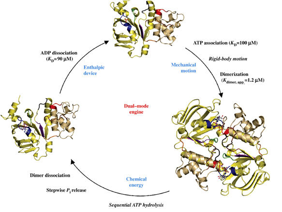
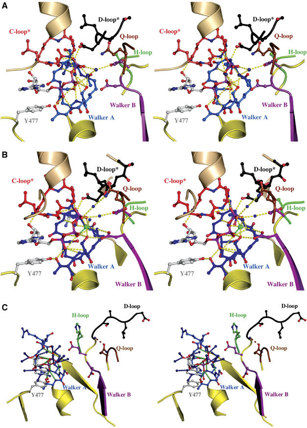
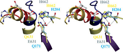
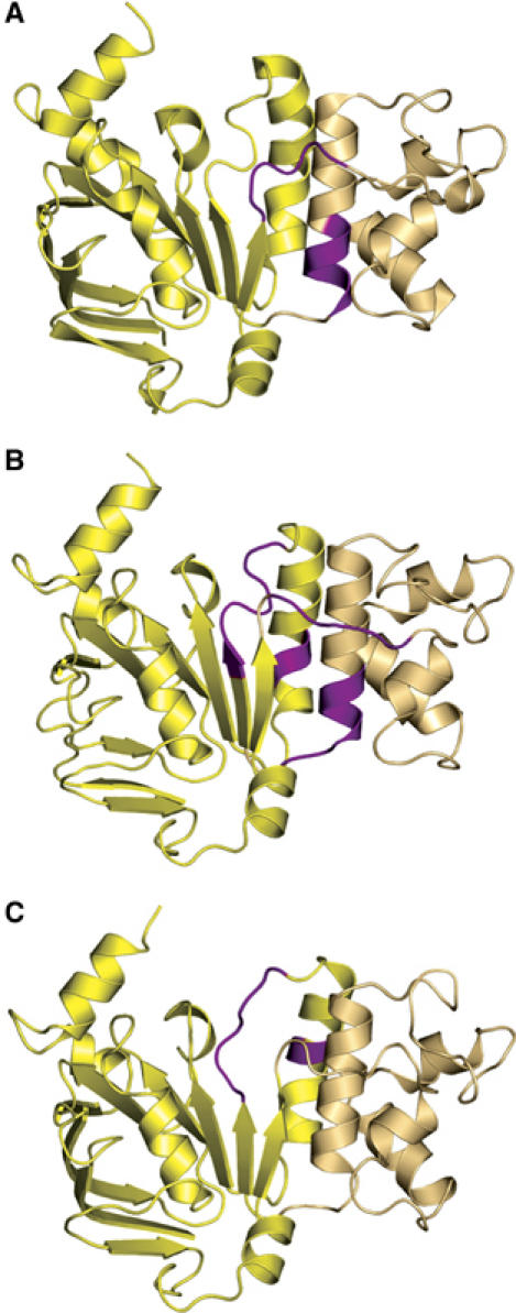
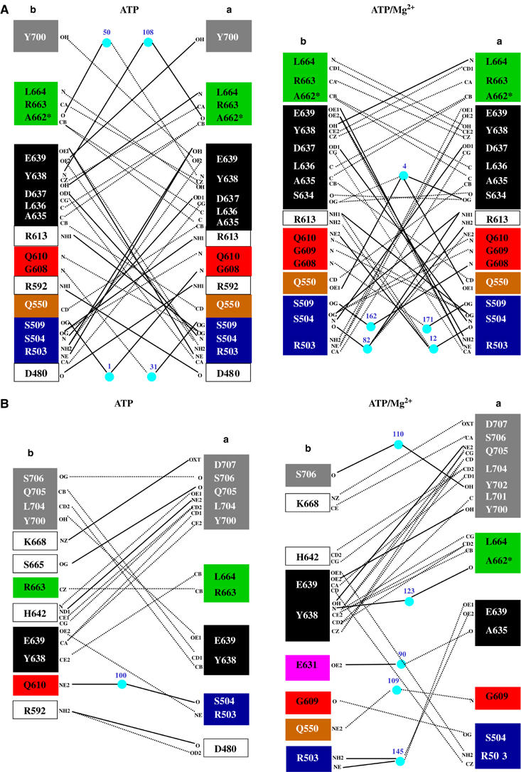
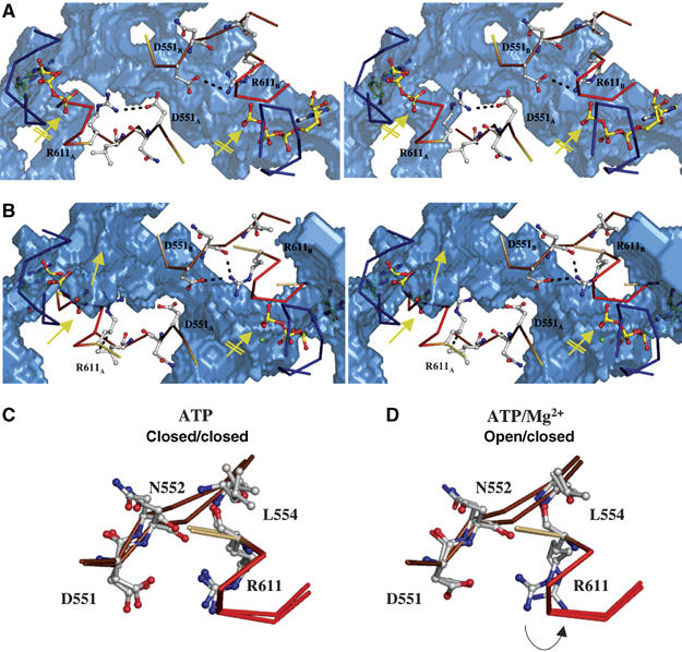
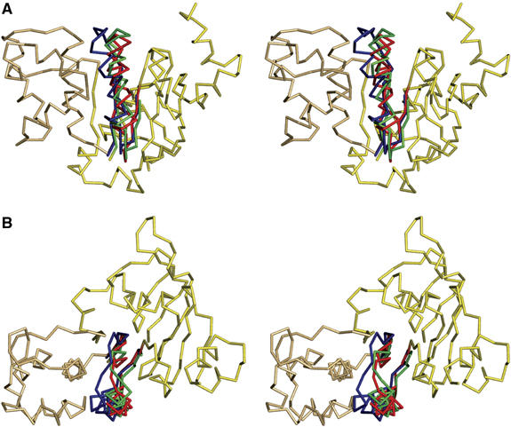
The ATP-binding cassette (ABC)-transporter haemolysin (Hly)B, a central element of a Type I secretion machinery, acts in concert with two additional proteins in Escherichia coli to translocate the toxin HlyA directly from the cytoplasm to the exterior. The basic set of crystal structures necessary to describe the catalytic cycle of the isolated HlyB-NBD (nucleotide-binding domain) has now been completed. This allowed a detailed analysis with respect to hinge regions, functionally important key residues and potential energy storage devices that revealed many novel features. These include a structural asymmetry within the ATP dimer that was significantly enhanced in the presence of Mg2+, indicating a possible functional asymmetry in the form of one open and one closed phosphate exit tunnel. Guided by the structural analysis, we identified two amino acids, closing one tunnel by an apparent salt bridge. Mutation of these residues abolished ATP-dependent cooperativity of the NBDs. The implications of these new findings for the coupling of ATP binding and hydrolysis to functional activity are discussed.


Figures

Figure 1
Figure 1
The catalytic cycle of the HlyB-NBD. Crystal structures of the monomeric nucleotide-free (Schmitt et al, 2003), dimeric ATP-bound (H662A and E631Q) and monomeric ADP-bound (wild type, E631Q and H662A) forms (this study) are shown. For simplicity, the structure of the ATP/Mg2+-bound form (Zaitseva et al, 2005a) is not shown. The catalytic domain is colored in light yellow and the helical domain in light tan. Conserved motifs are highlighted and color-coded as follows: Walker A (residues 502–510, blue), Q-loop (residues 549–556, brown), ABC-signature motif (residues 606–610, red), Pro-loop (residues 623–625, orange), Walker B (residues 626–630, magenta), D-loop (residues 634–637, black) and H-loop (residues 661–663, green). Bound ligands are shown in ball-and-stick representation. KD values were taken from Zaitseva et al (2005b).
Figure 2
Figure 2
Nucleotide-binding sites. Stereoview of the ATP-binding (A) and ATP/Mg2+-binding(B) sites. Color-coding is identical to Figure 1. Direct and water-mediated protein–ATP interactions are highlighted in yellow. Water molecules are shown as blue spheres and Mg2+ as a green sphere. The interaction between D637 of the D-loop of the trans monomer and S504 of the Walker A motif of the cis monomer is indicated. ATP and amino acids involved in ligand interactions are shown in ball-and-stick representation. * indicates conserved motifs of the trans monomer participating in ATP coordination. (C) Stereoview of the ADP-binding site. ADP and residues involved in ligand interactions are shown in ball-and-stick representation, water molecules are blue spheres, protein–ADP interactions are highlighted in green and ADP–water interactions in blue. Color-coding is identical to Figure 1. The interaction between the side chain of Q550 and the amide backbone of T633 is highlighted by a dashed, brown line.
Figure 3
Figure 3
Structural flexibility of the H662–E631 interaction. Stereoview of the ATP-binding site of the HlyB-NBD E631Q mutant, the E171Q mutant of MJ 0796 (PDB entry 1L2T) and the hypothetical model of the wild-type HlyB-NBD in complex with ATP. Side chains of the E631Q mutant of HlyB-NBD (Q631 and H662) are shown in yellow, side chains of the E171Q mutant of MJ0796 (Q171 and H204) in cyan and side chains of the model of wild-type HlyB-NBD (E631 and H662) in gray. The single interaction of Q631 with the side chain of H662 is highlighted in yellow; the bidentate interaction of H662 and E631 proposed in the linchpin model is highlighted in gray. Color-coding is identical to Figure 1.
Figure 4
Figure 4
Hinges and bends during functional transitions. Hinges involved in (A) the transition from the nucleotide-free to the ATP-bound state, (B) the transition from the ATP/Mg2+- to the ADP-bound state and (C) the transition from the ADP-bound to the nucleotide-free state. Hinge regions are highlighted in magenta. The catalytic domain is shown in light yellow and the helical domain in light tan. Structures shown correspond to the starting point of the functional transitions.
Figure 5
Figure 5
Importance of the cofactor Mg2+ in monomer–monomer interactions in the H662A dimer of HlyB. (A) Symmetric and (B) asymmetric interactions between the monomers of the ATP- (left panel) and ATP/Mg2+- (right panel) bound complexes of the HlyB-NBD H662A. Color-coding is identical to Figure 1. Solid lines represent hydrogen bonds (distance cutoff of 3.2 Å) and dashed line van der Waals interactions (distance cutoff of 4.0 Å). Light blue spheres represent water molecules. Letters indicate the atoms involved in the interactions.
Figure 6
Figure 6
An exit tunnel for inorganic phosphate. Stereoview of the ‘phosphate tunnel' within the two ATP-binding sites of the ATP-bound (A) and the ATP/Mg2+-bound (B) composite dimer of HlyB-NBD H662A. Color-coding is identical to Figure 1. For simplicity only the Walker A, Q-loop and ABC-signature motif are shown in ribbon. Mg2+ is shown as a green sphere and amino acids involved in interactions in ball-and-stick representation. The subscripts indicate the monomer to which the amino acids belong. The accessible solvent area (ASA) of the HlyB-NBD dimer is presented as a transparent, blue solid. The interaction between D551 and R611, which acts as a gate for tunnel opening (no interaction between D551 and R611, closest distance of 4.7 Å) and closing (interaction distance of 3.3 Å), is highlighted. The arrows in panels A and B indicate the open (standard arrow) and closed (crossed arrow) phosphate tunnel. Structural superimposition of the individual monomers of the ATP (C) and ATP/Mg2+ (D) dimer of the HlyB-NBD H662A mutant. The flip of R611 in the ATP/Mg2+ complex, which acts as a tunnel gate, is indicated by an arrow.
Figure 7
Figure 7
An enthalpic storage device for the chemical energy of ATP. (A) Structural superimposition of the nucleotide-free and ATP- and ADP-bound forms. For simplicity, only the structure of the ATP-bound form is shown as ribbons. Helix 6 of the nucleotide-free state is shown in green, for the ATP-bound state in red and for the ADP-bound state in blue. (B) 90° rotation in the plane with respect to panel A.

Similar articles

Cited by

References

    1. Chang G (2003) Structure of MsbA from Vibrio cholera: a multidrug resistance ABC transporter homolog in a closed conformation. J Mol Biol 330: 419–430 - PubMed
    1. Chang G, Roth CB (2001) Structure of MsbA from E. coli: a homolog of the multidrug resistance ATP binding cassette (ABC) transporters. Science 293: 1793–1800 - PubMed
    1. Chen J, Lu G, Lin J, Davidson AL, Quiocho FA (2003) A tweezers-like motion of the ATP-binding cassette dimer in an ABC transport cycle. Mol Cell 12: 651–661 - PubMed
    1. Chen J, Sharma S, Quiocho FA, Davidson AL (2001) Trapping the transition state of an ATP-binding cassette transporter: evidence for a concerted mechanism of maltose transport. Proc Natl Acad Sci USA 98: 1525–1530 - PMC - PubMed
    1. Davidson AL, Chen J (2004) ATP-binding cassette transporters in bacteria. Annu Rev Biochem 73: 241–268 - PubMed

Publication types

MeSH terms