Comprehensive analysis of myeloid lineage conversion using mice expressing an inducible form of C/EBP alpha
- PMID: 16858416
- PMCID: PMC1523173
- DOI: 10.1038/sj.emboj.7601199
Comprehensive analysis of myeloid lineage conversion using mice expressing an inducible form of C/EBP alpha
Abstract
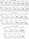
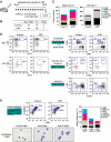
CCAAT/enhancer-binding protein (C/EBP) alpha is a critical regulator for early myeloid differentiation. Although C/EBPalpha has been shown to convert B cells into myeloid lineage, precise roles of C/EBPalpha in various hematopoietic progenitors and stem cells still remain obscure. To examine the consequence of C/EBPalpha activation in various progenitors and to address the underlying mechanism of lineage conversion in detail, we established transgenic mice expressing a conditional form of C/EBPalpha. Using these mice, we show that megakaryocyte/erythroid progenitors (MEPs) and common lymphoid progenitors (CLPs) could be redirected to functional macrophages in vitro by a short-term activation of C/EBPalpha, and the conversion occurred clonally through biphenotypic intermediate cells. Moreover, in vivo activation of C/EBPalpha in mice led to the increase of mature granulocytes and myeloid progenitors with a concomitant decrease of hematopoietic stem cells and nonmyeloid progenitors. Our study reveals that C/EBPalpha can activate the latent myeloid differentiation program of MEP and CLP and shows that its global activation affects multilineage homeostasis in vivo.
Figures

Similar articles
-
Gene coexpression analysis in single cells indicates lymphomyeloid copriming in short-term hematopoietic stem cells and multipotent progenitors.J Immunol. 2010 May 1;184(9):4907-17. doi: 10.4049/jimmunol.0902184. Epub 2010 Apr 5. J Immunol. 2010. PMID: 20368277
-
Establishment of the acute myeloid leukemia cell line Kasumi-6 from a patient with a dominant-negative mutation in the DNA-binding region of the C/EBPalpha gene.Genes Chromosomes Cancer. 2003 Feb;36(2):167-74. doi: 10.1002/gcc.10161. Genes Chromosomes Cancer. 2003. PMID: 12508245
-
Adult T-cell progenitors retain myeloid potential.Nature. 2008 Apr 10;452(7188):768-72. doi: 10.1038/nature06839. Nature. 2008. PMID: 18401412
-
[The hematopoietic stem cell and the stromal microenvironment].Therapie. 2001 Jul-Aug;56(4):383-4. Therapie. 2001. PMID: 11677858 Review. French.
-
Runx1, c-Myb, and C/EBPalpha couple differentiation to proliferation or growth arrest during hematopoiesis.J Cell Biochem. 2002;86(4):624-9. doi: 10.1002/jcb.10271. J Cell Biochem. 2002. PMID: 12210729 Review.
Cited by
-
C/EBPα in normal and malignant myelopoiesis.Int J Hematol. 2015 Apr;101(4):330-41. doi: 10.1007/s12185-015-1764-6. Epub 2015 Mar 10. Int J Hematol. 2015. PMID: 25753223 Free PMC article. Review.
-
Myeloid transformation of plasma cell myeloma: molecular evidence of clonal evolution revealed by next generation sequencing.Diagn Pathol. 2018 Feb 20;13(1):15. doi: 10.1186/s13000-018-0692-1. Diagn Pathol. 2018. PMID: 29463311 Free PMC article.
-
The complex landscape of haematopoietic lineage commitments is encoded in the coarse-grained endogenous network.R Soc Open Sci. 2021 Nov 3;8(11):211289. doi: 10.1098/rsos.211289. eCollection 2021 Nov. R Soc Open Sci. 2021. PMID: 34737882 Free PMC article.
-
Impaired hematopoietic differentiation of RUNX1-mutated induced pluripotent stem cells derived from FPD/AML patients.Leukemia. 2014 Dec;28(12):2344-54. doi: 10.1038/leu.2014.136. Epub 2014 Apr 15. Leukemia. 2014. PMID: 24732596
-
Zbtb1 prevents default myeloid differentiation of lymphoid-primed multipotent progenitors.Oncotarget. 2016 Sep 13;7(37):58768-58778. doi: 10.18632/oncotarget.11356. Oncotarget. 2016. PMID: 27542215 Free PMC article.
References
-
- Becker-Herman S, Lantner F, Shachar I (2002) Id2 negatively regulates B cell differentiation in the spleen. J Immunol 168: 5507–5513 - PubMed
-
- Busslinger M (2004) Transcriptional control of early B cell development. Annu Rev Immunol 22: 55–79 - PubMed
-
- Domen J, Gandy KL, Weissman IL (1998) Systemic overexpression of BCL-2 in the hematopoietic system protects transgenic mice from the consequences of lethal irradiation. Blood 91: 2272–2282 - PubMed
-
- Heath V, Suh HC, Holman M, Renn K, Gooya JM, Parkin S, Klarmann KD, Ortiz M, Johnson P, Keller J (2004) C/EBPalpha deficiency results in hyperproliferation of hematopoietic progenitor cells and disrupts macrophage development in vitro and in vivo. Blood 104: 1639–1647 - PubMed
Publication types
MeSH terms
Substances
LinkOut - more resources
Full Text Sources
Molecular Biology Databases
Miscellaneous

