

Olfactory mucosa autografts in human spinal cord injury: a pilot clinical study
- PMID: 16859223
- PMCID: PMC1864811
- DOI: 10.1080/10790268.2006.11753874
Olfactory mucosa autografts in human spinal cord injury: a pilot clinical study
Abstract
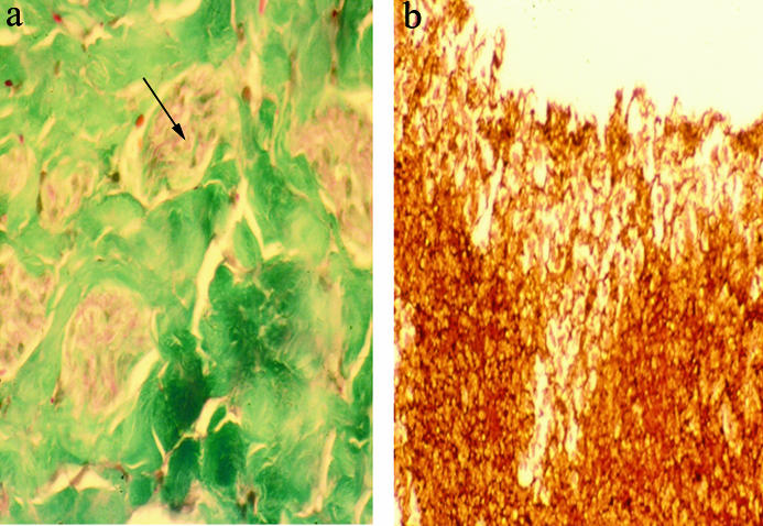
Background/objective: Olfactory mucosa is a readily accessible source of olfactory ensheathing and stem-like progenitor cells for neural repair. To determine the safety and feasibility of transplanting olfactory mucosa autografts into patients with traumatically injured spinal cords, a human pilot clinical study was conducted.
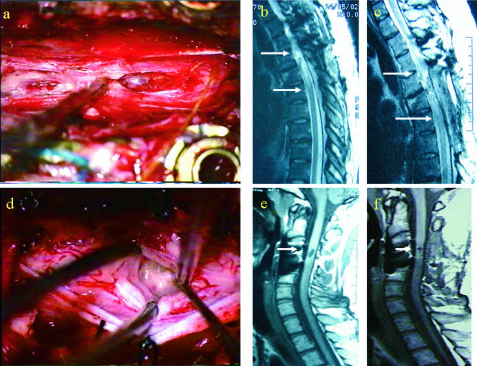
Methods: Seven patients ranging from 18 to 32 years of age (American Spinal Injury Association [ASIA] class A) were treated at 6 months to 6.5 years after injury. Olfactory mucosa autografts were transplanted into lesions ranging from 1 to 6 cm that were present at C4-T6 neurological levels. Operations were performed from July 2001 through March 2003. Magnetic resonance imaging (MRI), electromyography (EMG), and ASIA neurological and otolaryngological evaluations were performed before and after surgery.
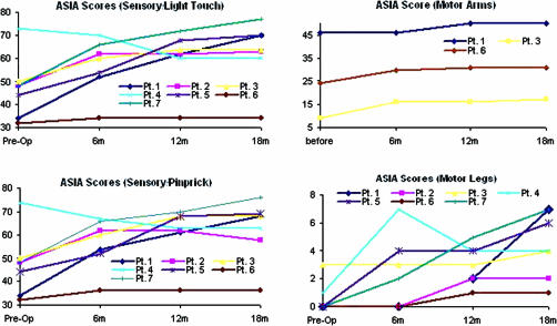
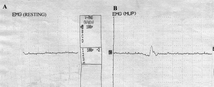
Results: MRI studies revealed moderate to complete filling of the lesion sites. Two patients reported return of sensation in their bladders, and one of these patients regained voluntary contraction of anal sphincter. Two of the 7 ASIA A patients became ASIA C. Every patient had improvement in ASIA motor scores. The mean increase for the 3 subjects with tetraplegia in the upper extremities was 6.3 +/- 1.2 (SEM), and the mean increase for the 4 subjects with paraplegia in the lower extremities was 3.9 +/- 1.0. Among the patients who improved in their ASIA sensory neurological scores (all except one patient), the mean increase was 20.3 +/- 5.0 for light touch and 19.7 +/- 4.6 for pinprick. Most of the recovered sensation below the initial level of injury was impaired. Adverse events included sensory decrease in one patient that was most likely caused by difficulty in locating the lesion, and there were a few instances of transient pain that was relieved by medication. EMG revealed motor unit potential when the patient was asked to perform movement.
Conclusion: This study shows that olfactory mucosa autograft transplantation into the human injured spinal cord is feasible, relatively safe, and potentially beneficial. The procedure involves risks generally associated with any surgical procedure. Long-term patient monitoring is necessary to rule out any delayed side effects and assess any further improvements.
Figures
Similar articles
-
Olfactory mucosal autografts and rehabilitation for chronic traumatic spinal cord injury.Neurorehabil Neural Repair. 2010 Jan;24(1):10-22. doi: 10.1177/1545968309347685. Epub 2009 Sep 30. Neurorehabil Neural Repair. 2010. PMID: 19794133 Clinical Trial.
-
Transplantation of autologous olfactory ensheathing cells in complete human spinal cord injury.Cell Transplant. 2013;22(9):1591-612. doi: 10.3727/096368912X663532. Cell Transplant. 2013. PMID: 24007776 Clinical Trial.
-
Autologous olfactory ensheathing cell transplantation in human paraplegia: a 3-year clinical trial.Brain. 2008 Sep;131(Pt 9):2376-86. doi: 10.1093/brain/awn173. Epub 2008 Aug 8. Brain. 2008. PMID: 18689435 Free PMC article. Clinical Trial.
-
Olfactory ensheathing cells from the nose: clinical application in human spinal cord injuries.Exp Neurol. 2011 May;229(1):174-80. doi: 10.1016/j.expneurol.2010.08.025. Epub 2010 Sep 9. Exp Neurol. 2011. PMID: 20832402 Review.
-
Olfactory ensheathing cells - another miracle cure for spinal cord injury?Nat Rev Neurosci. 2001 May;2(5):369-75. doi: 10.1038/35072576. Nat Rev Neurosci. 2001. PMID: 11331921 Review.
Cited by
-
Differentiation of CD133+ stem cells from amyotrophic lateral sclerosis patients into preneuron cells.Stem Cells Transl Med. 2013 Feb;2(2):129-35. doi: 10.5966/sctm.2012-0077. Epub 2013 Jan 22. Stem Cells Transl Med. 2013. PMID: 23341441 Free PMC article.
-
Curiosity and cure: translational research strategies for neural repair-mediated rehabilitation.Dev Neurobiol. 2007 Aug;67(9):1133-47. doi: 10.1002/dneu.20514. Dev Neurobiol. 2007. PMID: 17514711 Free PMC article. Review.
-
Olfactory Ensheathing Cells for Spinal Cord Injury: Sniffing Out the Issues.Cell Transplant. 2018 Jun;27(6):879-889. doi: 10.1177/0963689718779353. Epub 2018 Jun 8. Cell Transplant. 2018. PMID: 29882418 Free PMC article. Review.
-
Motor evoked potential and voluntary EMG activity after olfactory mucosal autograft transplantation in a case of chronic, complete spinal cord injury: case report.Spinal Cord Ser Cases. 2016 Jan 7;2:15018. doi: 10.1038/scsandc.2015.18. eCollection 2016. Spinal Cord Ser Cases. 2016. PMID: 28053727 Free PMC article.
-
Cell Therapy From Bench to Bedside Translation in CNS Neurorestoratology Era.Cell Med. 2010 Jan 1;1(1):15-46. doi: 10.3727/215517910X516673. Cell Med. 2010. PMID: 21359168 Free PMC article.
References
-
- McDonald JW, Liu XZ, Qu Y, et al. Transplanted embryonic stem cells survive, differentiate and promote recovery in injured rat spinal cord. Nat Med. 1999;5:1410–1412. - PubMed
-
- Akiyama Y, Honmou O, Kato T, Uede T, Hashi K, Kocsis JD. Transplantation of clonal neural precursor cells derived from adult human brain establishes functional peripheral myelin in the rat spinal cord. Exp Neurol. 2001;167:27–39. - PubMed
-
- Reier PJ, Houle JD, Jakeman L, Winialski D, Tessler A. Transplantation of fetal spinal cord tissue into acute and chronic hemisection and contusion lesions of the adult rat spinal cord. Prog Brain Res. 1988;78:173–179. - PubMed
-
- Xu XM, Zhang SX, Li H, Aebischer P, Bunge MB. Regrowth of axons into the distal spinal cord through a Schwann-cell-seeded mini-channel implanted into hemisected adult rat spinal cord. Eur J Neurosci. 1999;11:1723–1740. - PubMed
-
- Grill RJ, Blesch A, Tuszynski MH. Robust growth of chronically injured spinal cord axons induced by grafts of genetically modified NGF-secreting cells. Exp Neurol. 1997;148:444–452. - PubMed
Publication types
MeSH terms
LinkOut - more resources
Full Text Sources
Other Literature Sources
Medical
Miscellaneous