. 2006 Oct 15;399(2):249-56.
doi: 10.1042/BJ20060781.

A protein extension to shorten RNA: elongated elongation factor-Tu recognizes the D-arm of T-armless tRNAs in nematode mitochondria

Affiliations

A protein extension to shorten RNA: elongated elongation factor-Tu recognizes the D-arm of T-armless tRNAs in nematode mitochondria

Masayuki Sakurai et al. Biochem J. .

Abstract

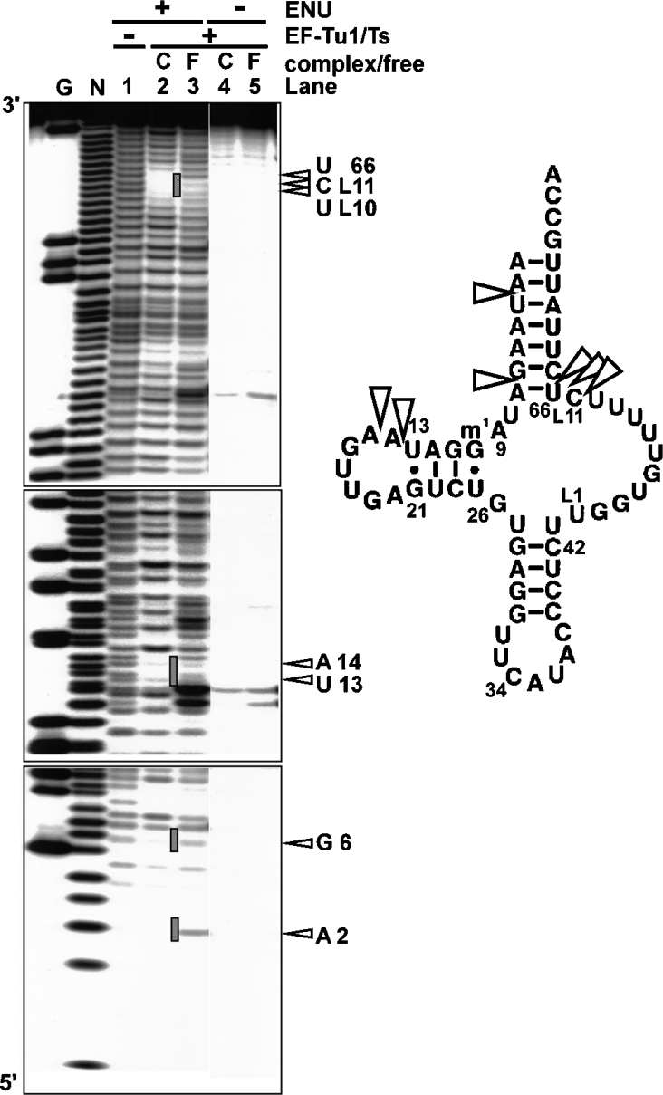
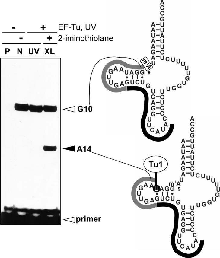
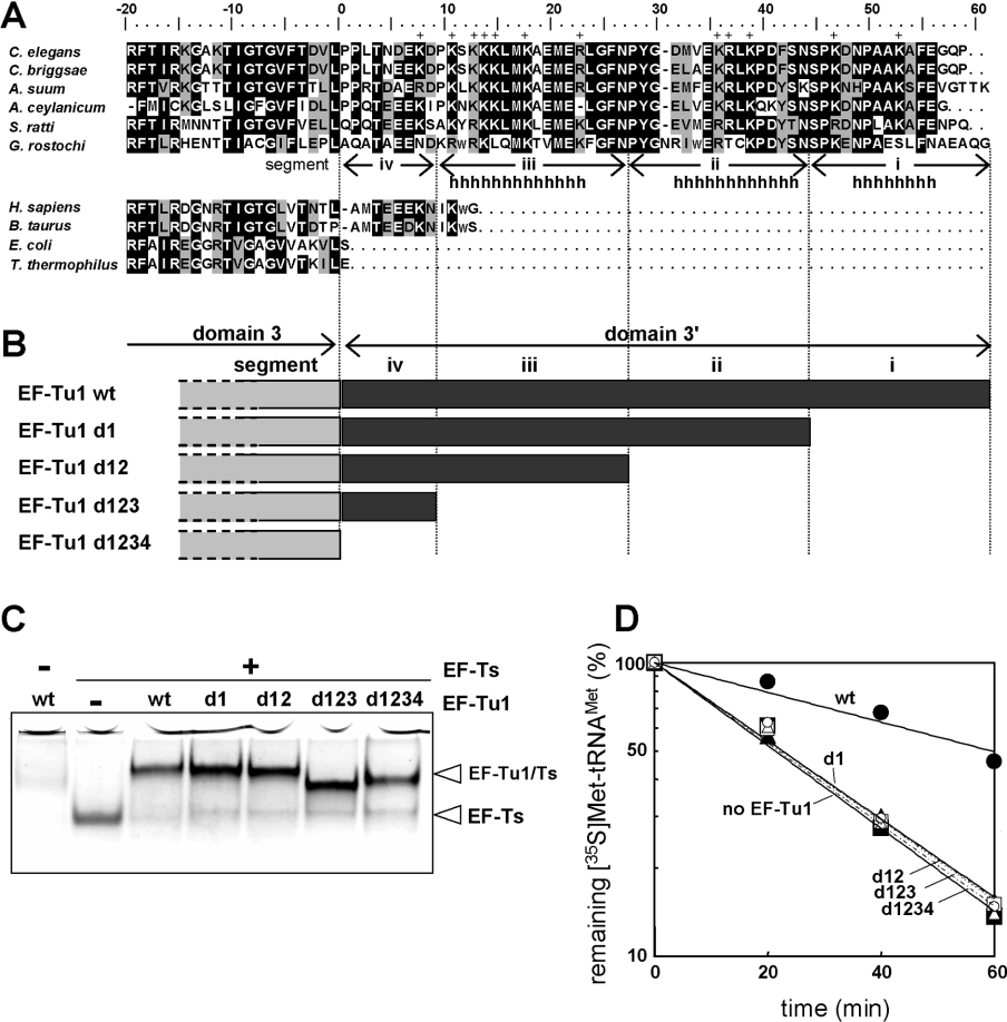
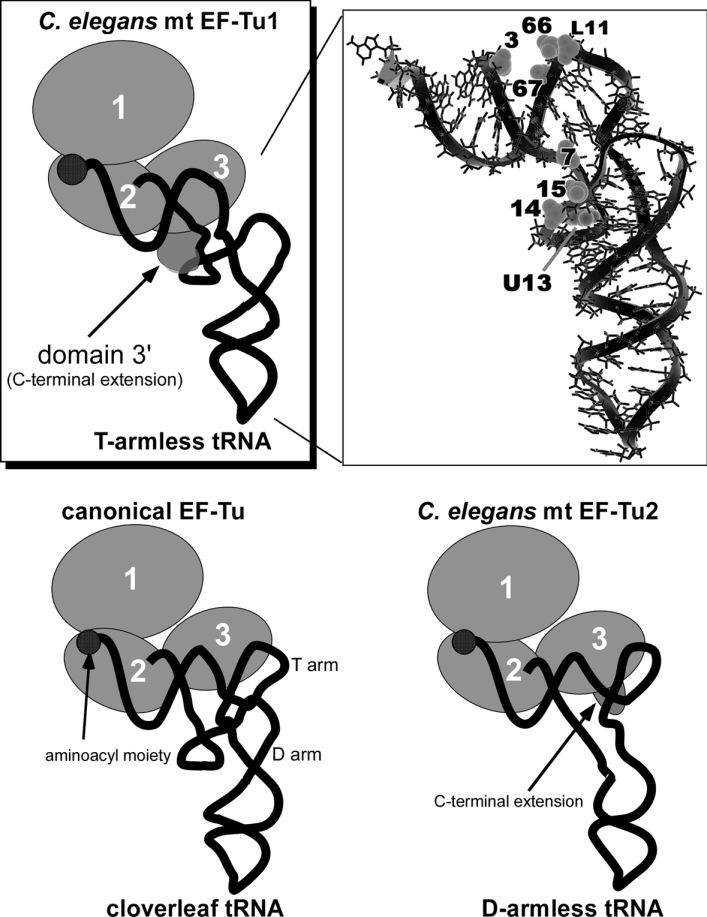
Nematode mitochondria possess extremely truncated tRNAs. Of 22 tRNAs, 20 lack the entire T-arm. The T-arm is necessary for the binding of canonical tRNAs and EF (elongation factor)-Tu (thermo-unstable). The nematode mitochondrial translation system employs two different EF-Tu factors named EF-Tu1 and EF-Tu2. Our previous study showed that nematode Caenorhabditis elegans EF-Tu1 binds specifically to T-armless tRNA. C. elegans EF-Tu1 has a 57-amino acid C-terminal extension that is absent from canonical EF-Tu, and the T-arm-binding residues of canonical EF-Tu are not conserved. In this study, the recognition mechanism of T-armless tRNA by EF-Tu1 was investigated. Both modification interference assays and primer extension analysis of cross-linked ternary complexes revealed that EF-Tu1 interacts not only with the tRNA acceptor stem but also with the D-arm. This is the first example of an EF-Tu recognizing the D-arm of a tRNA. The binding activity of EF-Tu1 was impaired by deletion of only 14 residues from the C-terminus, indicating that the C-terminus of EF-Tu1 is required for its binding to T-armless tRNA. These results suggest that C. elegans EF-Tu1 recognizes the D-arm instead of the T-arm by a mechanism involving its C-terminal region. This study sheds light on the co-evolution of RNA and RNA-binding proteins in nematode mitochondria.


Figures

Figure 1
Figure 1. Identification of the EF-Tu-binding sites on A. suum mt tRNAMet by the ENU-modification interference assay
Left-hand side, autoradiographs of the ENU-modification interference assay using C. elegans mt EF-Tu1. Lanes: G, RNase T1 ladder; N, alkaline ladder; lanes 1–3, ENU-modified Val-tRNA; lanes 4 and 5, Val-tRNA without ENU modification; lanes 2, 3, 4 and 5, the Val-tRNA incubated with EF-Tu1–EF-Ts; lanes 2 and 4, the Val-tRNA in the ternary complex; lanes 3 and 5, the Val-tRNA free from EF-Tu. The open triangles and hatched lines show the ENU-modification interference of the complex formation. Right-hand side, the EF-Tu1-binding sites on the tRNAMet. The open triangles indicate phosphates interacting with the EF-Tu1. The base numbering is according to [10].
Figure 2
Figure 2. Primer extension analysis of cross-linked ternary complexes using a 5′-32P-labelled DNA primer (Primer 1)
Left-hand side, A. suum mt Val-tRNAMet (m1A9; lane N), the ternary complex irradiated by UV (lane UV) and the ternary complex irradiated by UV in the presence of 2-iminothiolane (lane XL) were analysed by primer extension on 15% denaturing PAGE. Lane P, primer only; lanes N, UV and XL, Val-tRNAMet was used. Right-hand side, diagrams of the primer extension results. Black and grey lines show Primer 1 complementarity to G21–A37 of the tRNA, and the extension of the primer respectively. The primer extensions were stopped at G10 in lanes N, UV and XL because of inhibition by the 1-methyl group at A9.
Figure 3
Figure 3. C-terminal deletion mutants of C. elegans EF-Tu1
(A) Amino acid alignment of EF-Tu1 homologues. EF-Tu1 sequences of C. elegans (accession number D38471), and its homologues in other nematodes, Caenorhabditis briggsae (CAE57516), A. suum (AB211994, the present study), Ancylostoma ceylanicum (CA341502, EST data), S. ratti (AB211996, the present study), and G. rostochiensis (AB211995, the present study). EF-Tu sequences of Homo sapiens (X84694), Bos taurus (L38996), E. coli (P02990) and T. thermophilus (SP07157) were also aligned. The alignment was carried out using Clustal X [51] followed by manual modifications. Black and gray areas indicate identical and similar amino acid sequences respectively. The positively charged amino acids in domain 3′ of C. elegans EF-Tu1 are indicated by ‘+’. The probable α-helix region of C. elegans mt EF-Tu1 predicted by a secondary structure prediction program is indicated by ‘h’ [32]. The predicted three helical regions are represented as segments i, ii and iii and the rest of domain 3′ is represented as segment iv. (B) Schematic representations of C-terminal deletion mutants of C. elegans EF-Tu1. (C) Native 9% PAGE analysis of complexes of EF-Tu1 deletion mutants and C. elegans mt EF-Ts. (D) The aa-tRNA-binding activities of the EF-Tu1 deletion mutants. A deacylation protection assay of the aminoacyl ester bond against hydrolysis of A. suum mt [35S]Met-tRNAMet (initial concentration 50 nM) was performed in the absence of EF-Tu (filled triangles and solid line) and in the presence of 5 μM C. elegans EF-Tu1–EF-Ts complex (filled circles and solid line) or mutant EF-Tu1–EF-Ts complex (d1, filled squares and solid line; d12, open circles and dotted line; d123, open squares and dotted line; d1234, open triangles and dotted line).
Figure 4
Figure 4. Models of the ternary complexes
EF-Tu is shown as a set of circles, each of which corresponds to the domain numbers. The tRNAs are portrayed as simplified backbones with the aminoacyl moiety at the 3′-terminus depicted by filled circles. GTP is not shown. The ternary complex including nematode mt EF-Tu1 and the T-armless tRNA (top left). The hatched domain shows the predicted location of the EF-Tu1 domain 3′. A. suum mt tRNAMet lacking the T-arm is presented by the structural model (top right) [52]. The phosphates important for EF-Tu1-binding, detected by modification interference (3, 7, 14, 15, L11, 66 and 67), and the base cross-linked with EF-Tu1 (U13) are shown in the space-filled representation. The tRNA chemical structure is shown with thin lines and its backbone with the bold line. The bacterial ternary complex is shown bottom left [2]. The ternary complex including nematode mt EF-Tu2 and D-armless Ser-tRNA is shown bottom right [20].

Comment in

References

    1. Sprinzl M. Elongation factor Tu: a regulatory GTPase with an integrated effector. Trends Biochem. Sci. 1994;19:245–250. - PubMed
    1. Nissen P., Kjeldgaard M., Thirup S., Polekhina G., Reshetnikova L., Clark B. F., Nyborg J. Crystal structure of the ternary complex of Phe-tRNAPhe, EF-Tu, and a GTP analog. Science. 1995;270:1464–1472. - PubMed
    1. Nissen P., Thirup S., Kjeldgaard M., Nyborg J. The crystal structure of Cys-tRNACys-EF-Tu-GDPNP reveals general and specific features in the ternary complex and in tRNA. Structure. 1999;7:143–156. - PubMed
    1. Clark B. F. C., Kjeldgaard M., Barciszewski J., Sprinzl M. Recognition of aminoacyl-tRNAs by protein elongation factors. tRNA: Structure, Biosynthesis, and Function. In: Soll D., RajBhandary U. L., editors. Washington, DC: American Society for Microbiology; 1995. pp. 423–442.
    1. Rudinger J., Blechschmidt B., Ribeiro S., Sprinzl M. Minimalist aminoacylated RNAs as efficient substrates for elongation factor Tu. Biochemistry. 1994;33:5682–5688. - PubMed

Publication types

MeSH terms

Associated data