. 2006 Dec 15;400(3):531-40.
doi: 10.1042/BJ20060874.

Directed evolution of a monomeric, bright and photostable version of Clavularia cyan fluorescent protein: structural characterization and applications in fluorescence imaging

Affiliations

Directed evolution of a monomeric, bright and photostable version of Clavularia cyan fluorescent protein: structural characterization and applications in fluorescence imaging

Hui-wang Ai et al. Biochem J. .

Abstract

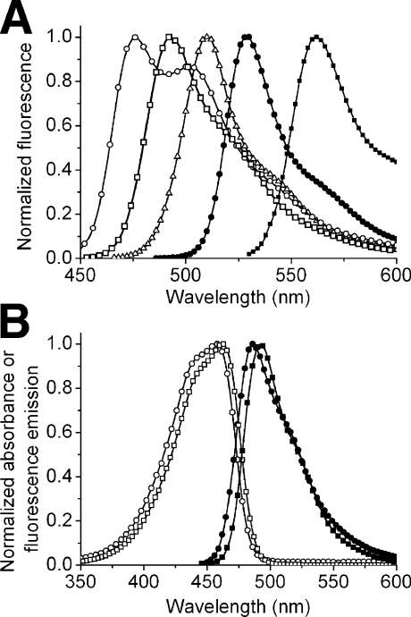
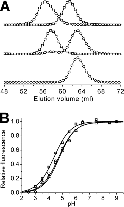
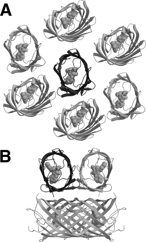
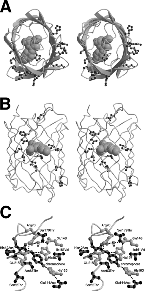
The arsenal of engineered variants of the GFP [green FP (fluorescent protein)] from Aequorea jellyfish provides researchers with a powerful set of tools for use in biochemical and cell biology research. The recent discovery of diverse FPs in Anthozoa coral species has provided protein engineers with an abundance of alternative progenitor FPs from which improved variants that complement or supersede existing Aequorea GFP variants could be derived. Here, we report the engineering of the first monomeric version of the tetrameric CFP (cyan FP) cFP484 from Clavularia coral. Starting from a designed synthetic gene library with mammalian codon preferences, we identified dimeric cFP484 variants with fluorescent brightness significantly greater than the wild-type protein. Following incorporation of dimer-breaking mutations and extensive directed evolution with selection for blue-shifted emission, high fluorescent brightness and photostability, we arrived at an optimized variant that we have named mTFP1 [monomeric TFP1 (teal FP 1)]. The new mTFP1 is one of the brightest and most photostable FPs reported to date. In addition, the fluorescence is insensitive to physiologically relevant pH changes and the fluorescence lifetime decay is best fitted as a single exponential. The 1.19 A crystal structure (1 A=0.1 nm) of mTFP1 confirms the monomeric structure and reveals an unusually distorted chromophore conformation. As we experimentally demonstrate, the high quantum yield of mTFP1 (0.85) makes it particularly suitable as a replacement for ECFP (enhanced CFP) or Cerulean as a FRET (fluorescence resonance energy transfer) donor to either a yellow or orange FP acceptor.


Figures

Figure 1
Figure 1. Absorbance and fluorescence emission spectra of a selection of FPs
(A) Fluorescence emission spectra of mCerulean (○) [14,34], mTFP1 (□), EGFP (△), Citrine (●) [32] and mOrange (■) [33]. (B) Absorbance (open symbols) and fluorescence emission (filled symbols) spectra of dTFP0.2 (○, ●) and mTFP1 (□, ■).
Figure 2
Figure 2. Characterization of oligomeric structure and pH-sensitivity
(A) Gel filtration chromatography elution profile of dTFP0.2 and mTFP1. Detection is at either 450 nm (○) or 550 nm (□). The upper profile is a co-injection of dTFP0.2 and monomeric mCherry [33], the middle profile is dimeric dTomato [33] and mTFP1, and the lower profile is mTFP1 alone. Earlier generations of mTFP had elution times identical with mTFP1 (results not shown). The small 450 nm peak at the dimer elution volume in the middle profile is due to the weak absorbance of dTomato at this wavelength. (B) pH-dependence of the fluorescence emission of mCerulean (○), mTFP1 (□) and mECFP (△).
Figure 3
Figure 3. Crystal packing of mTFP1 and amFP486
(A) Shown in dark grey cartoon representation is the asymmetric unit of mTFP1 (a single polypeptide) crystallized in the monoclinic space group C2. Also shown are the proximal copies of the protein (light grey cartoon) as generated using crystallographic symmetry operations. As expected for a monomeric variant, none of the proximal protein subunits pack in orientations similar to those typically observed in Anthozoa FPs such as the highly homologous amFP486 [48] shown in (B). (B) For amFP486 crystallized in the tetragonal space group I4122 (Protein Data Bank code 2A46), the asymmetric unit contains a single polypeptide (dark grey cartoon), which is shown in the same orientation as mTFP1 in (A) [48]. The additional three subunits (light grey cartoon) of the tightly packed tetrameric biological unit were generated using the crystallographic symmetry operations.
Figure 4
Figure 4. Location of beneficial mutations in mTFP1
(A) Stereoview of mTFP1 with the polypeptide backbone in cartoon representation and the chromophore in space filling representation. All residues with fully solvent exposed side chains that are mutated relative to the wild-type cFP484 are shown in ball-and-stick representation. The protein is viewed from the same orientation as the single asymmetric unit shown in Figures 3(A) and 3(B). By comparison with the amFP486 structure shown in Figure 3(B), it is apparent that the external mutations are clustered in the former protein–protein interfaces of the tetrameric wild-type protein. (B) Stereoview of mTFP1 with all residues that are directed towards the interior of the β-barrel and mutated relative to cFP484 shown in ball-and-stick representation. (C) Stereoview of the chromophore region of mTFP1 showing the chromophore (black bonds with grey atoms), all mutations in the immediate vicinity of the chromophore (grey bonds with black atoms), and a selection of residues (i.e. the quadrupole salt-bridge network and His163) that were not mutated but are discussed in the text (white bonds with grey atoms).
Figure 5
Figure 5. mTFP1 as a FRET donor to a YFP or mOrange acceptor
(A) Shown are the in vitro emission spectra of mTFP1–YC3.3 [13] with no Ca2+ (○), 10 mM Ca2+ (□) and no FRET (△). To obtain the ‘no FRET’ spectrum, the linker between the two FPs was digested with trypsin under conditions where the FPs remain intact. Shown in grey lines is a plot of percentage transmittance versus wavelength for the custom filter set used for imaging of mTFP1–YFP FRET to produce the results shown in Figure 7. Filters are designated with Chroma Technology part numbers. (B) Shown are the in vitro emission spectra for an mTFP1–mOrange fusion protein before (○), and after (△), proteolysis of the linker peptide.
Figure 6
Figure 6. Localization of mTFP1 fusion proteins in live HeLa cells
Confocal fluorescence images of HeLa cells expressing either mTFP1–β-actin (A) or mTFP1–α-tubulin (B). Scale bar, 10 μm in both images.
Figure 7
Figure 7. FRET imaging of mTFP1 with a YFP acceptor
Shown is a plot of the emission ratio versus time for three HeLa cells (see donor fluorescence image in the inset) expressing mTFP1–YC3.3 targeted to the ER. Images in both the donor and acceptor emission channels were acquired every 5 s with identical exposure times (300 ms) and 10% ND filters. Histamine (200 μM) or ionomycin (2 μM) was added at the indicated time points.

References

    1. Tsien R. Y. The green fluorescent protein. Annu. Rev. Biochem. 1998;67:509–544. - PubMed
    1. Matz M. V., Fradkov A. F., Labas Y. A., Savitsky A. P., Zaraisky A. G., Markelov M. L., Lukyanov S. A. Fluorescent proteins from nonbioluminescent Anthozoa species. Nat. Biotechnol. 1999;17:969–973. - PubMed
    1. Verkhusha V. V., Lukyanov K. A. The molecular properties and applications of Anthozoa fluorescent proteins and chromoproteins. Nat. Biotechnol. 2004;22:289–296. - PubMed
    1. Zhang J., Campbell R. E., Ting A. Y., Tsien R. Y. Creating new fluorescent probes for cell biology. Nat. Rev. Mol. Cell Biol. 2002;3:906–918. - PubMed
    1. Chudakov D. M., Lukyanov S., Lukyanov K. A. Fluorescent proteins as a toolkit for in vivo imaging. Trends Biotechnol. 2005;23:605–613. - PubMed

Publication types

Substances