. 2006;7(7):R60.
doi: 10.1186/gb-2006-7-7-r60.

The prokaryotic antecedents of the ubiquitin-signaling system and the early evolution of ubiquitin-like beta-grasp domains

Affiliations

The prokaryotic antecedents of the ubiquitin-signaling system and the early evolution of ubiquitin-like beta-grasp domains

Lakshminarayan M Iyer et al. Genome Biol. 2006.

Abstract

Background: Ubiquitin (Ub)-mediated signaling is one of the hallmarks of all eukaryotes. Prokaryotic homologs of Ub (ThiS and MoaD) and E1 ligases have been studied in relation to sulfur incorporation reactions in thiamine and molybdenum/tungsten cofactor biosynthesis. However, there is no evidence for entire protein modification systems with Ub-like proteins and deconjugation by deubiquitinating enzymes in prokaryotes. Hence, the evolutionary assembly of the eukaryotic Ub-signaling apparatus remains unclear.

Results: We systematically analyzed prokaryotic Ub-related beta-grasp fold proteins using sensitive sequence profile searches and structural analysis. Consequently, we identified novel Ub-related proteins beyond the characterized ThiS, MoaD, TGS, and YukD domains. To understand their functional associations, we sought and recovered several conserved gene neighborhoods and domain architectures. These included novel associations involving diverse sulfur metabolism proteins, siderophore biosynthesis and the gene encoding the transfer mRNA binding protein SmpB, as well as domain fusions between Ub-like domains and PIN-domain related RNAses. Most strikingly, we found conserved gene neighborhoods in phylogenetically diverse bacteria combining genes for JAB domains (the primary de-ubiquitinating isopeptidases of the proteasomal complex), along with E1-like adenylating enzymes and different Ub-related proteins. Further sequence analysis of other conserved genes in these neighborhoods revealed several Ub-conjugating enzyme/E2-ligase related proteins. Genes for an Ub-like protein and a JAB domain peptidase were also found in the tail assembly gene cluster of certain caudate bacteriophages.

Conclusion: These observations imply that members of the Ub family had already formed strong functional associations with E1-like proteins, UBC/E2-related proteins, and JAB peptidases in the bacteria. Several of these Ub-like proteins and the associated protein families are likely to function together in signaling systems just as in eukaryotes.


Figures

Figure 1
Figure 1
ThiS/MoaD/Ubiquitin-based protein conjugation system. The figure shows different themes by which a ThiS/MoaD/Ubiquitin-like polypeptide participates in thiamine biosynthesis, MoCo/WCo biosynthesis, and the ubiquitin conjugation/deconjugation system and the siderophore biosynthesis pathways. The '?' refers to the speculated part of the pathway inferred from operon organization. SUB refers to the polypeptide/protein substrate.
Figure 2
Figure 2
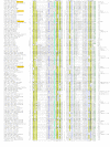
Multiple alignment of ThiS/MoaD-like ubiquitin domain containing proteins. Proteins are listed by gene name, species abbreviation and gi number, separated by underscores. Amino acid residues are colored according to side chain properties and the extent of conservation in the multiple alignment. Coloring is indicative of 70% consensus, which is shown on the last line of the alignment. Consensus similarity designations and coloring scheme are as follows: h, hydrophobic residues (ACFILMVWY), shaded yellow; s, small residues (AGSVCDN), colored green; o, alcohol group containing residues (ST), colored blue; and b, big residues (EFHIKLMQRWY), colored purple and shaded in light gray. Secondary structure assignments are shown above the alignment, where E represents a strand and H represents a helix. The families of the ubiquitin-related domains are shown to the right. Also shown to the right are the row numbers in Table 1, which describe a particular family. Species abbreviations are as follows: Aaeo, Aquifex aeolicus; Adeh, Anaeromyxobacter dehalogenans; Aehr, Alkalilimnicola ehrlichei; Aful, Archaeoglobus fulgidus; Amac, Alteromonas macleodii; Amet, Alkaliphilus metalliredigenes; Asp., Arthrobacter sp.; Azsp, Azoarcus sp.; Atha, Arabidopsis thaliana; Avar, Anabaena variabilis; BJK0, Bacteriophage JK06; Bbro, Bordetella bronchiseptica; Bcen, Burkholderia cenocepacia; Bcep, Burkholderia cepacia; Bcer, Bacillus cereus; Bcla, Bacillus clausii; Blic, Bacillus licheniformis, Bphi, Bacteriophage phiE125; Bsp., Bradyrhizobium sp.; Bsub, Bacillus subtilis; Bthe, Bacteroides thetaiotaomicron; Bthu, Bacillus thuringiensis; Bvie, Burkholderia vietnamiensis; Cace, Clostridium acetobutylicum; Caur, Chloroflexus aurantiacus; Ccol, Campylobacter coli; Cele, Caenorhabditis elegans; Cinc, Chlamydomonas incerta; Cjej, Campylobacter jejuni; Cnec, Cupriavidus necator; Cper, Clostridium perfringens; Cpha, Chlorobium phaeobacteroides; Csac, Caldicellulosiruptor saccharolyticus; Ctet, Clostridium tetani; Dace, Desulfuromonas acetoxidans; Daro, Dechloromonas aromatica; Dhaf, Desulfitobacterium hafniense; Dmel, Drosophila melanogaster; Dpsy, Desulfotalea psychrophila; Drad, Deinococcus radiodurans; Dvul, Desulfovibrio vulgaris; Ecol, Escherichia coli; Elit, Erythrobacter litoralis; Epha, Enterobacteria phage; Fsp., Frankia sp.; Glam, Giardia lamblia; Gmet, Geobacter metallireducens; Goxy, Gluconobacter oxydans; Gsul, Geobacter sulfurreducens; Gura, Geobacter uraniumreducens; Hsap, Homo sapiens; Hsp., Halobacterium sp.; Mace, Methanosarcina acetivorans; Maqu, Marinobacter aquaeolei; Mdeg, Microbulbifer degradans; Mfla, Mycobacterium flavescens, Mgry, Magnetospirillum gryphiswaldense; Mjan, Methanocaldococcus jannaschii; Mlot, Mesorhizobium loti; Mmag, Magnetospirillum magnetotacticum; Mmus, Mus musculus; Msp., Magnetococcus sp.; Mtub, Mycobacterium tuberculosis; Neur, Nitrosomonas europaea; Nfar, Nocardia farcinica; Nham, Nitrobacter hamburgensis; Nisp, Nitrobacter sp.; Nmen, Neisseria meningitidis; Nmul, Nitrosospira multiformis; Noce, Nitrosococcus oceani; Nosp, Nocardioides sp.; Nsp., Nostoc sp.; Nwin, Nitrobacter winogradskyi; Obat, Oceanicola batsensis; PBP-, Phage BP-4795; Paby, Pyrococcus abyssi; Paer, Pseudomonas aeruginosa; Parc, Psychrobacter arcticus; Pber, Parvularcula bermudensis; Pcar, Pelobacter carbinolicus; Pflu, Pseudomonas fluorescens; Pfur, Pyrococcus furiosus; Phor, Pyrococcus horikoshii; Pmen, Pseudomonas mendocina; Pnap, Polaromonas naphthalenivorans; Posp, Polaromonas sp.; Ppro, Pelobacter propionicus; Pput, Pseudomonas putida; Psp., Pseudomonas sp.; Psyr, Pseudomonas syringae; Retl, Rhizobium etli; Reut, Ralstonia eutropha; Rfer, Rhodoferax ferrireducens; Rmet, Ralstonia metallidurans; Rosp, Roseovarius sp.; Rpal, Rhodopseudomonas palustris; Rsol, Ralstonia solanacearum; RhNGR234a, Rhizobium sp. NGR234a plasmid; Rsp, Rhizobium sp. NGR234; Rsph, Rhodobacter sphaeroides; Rusp, Ruegeria sp.; Rxyl, Rubrobacter xylanophilus; Saci, Syntrophus aciditrophicus; Save, Streptomyces avermitilis; Scer, Saccharomyces cerevisiae; Scoe, Streptomyces coelicolor; Sdis, Spisula solidissima; Sepi, Staphylococcus epidermidis; Spom, Schizosaccharomyces pombe; Spur, Strongylocentrotus purpuratus; Srub, Salinibacter ruber; Ssol, Sulfolobus solfataricus; Ssp., Synechocystis sp.; Swsp, Shewanella sp.; Tfus, Thermobifida fusca; Tmar, Thermotoga maritima; Tpar, Theileria parva; Vcho, Vibrio cholerae; Vfis, Vibrio fischeri; Vpar, Vibrio parahaemolyticus; Vsp., Vibrio sp.; Wsuc, Wolinella succinogenes; Xaxo, Xanthomonas axonopodis; Xcam, Xanthomonas campestris; Ymol, Yersinia mollaretii; Ypes, Yersinia pestis.
Figure 3
Figure 3
Domain architectures of ThiS/MoaD-like ubiquitin domains and functionally associated proteins. Architectures belonging to a particular gene neighborhood or related pathway are grouped in boxes. Proteins are identified below the architectures by gene name, species abbreviation and gi number, demarcated by underscores. Proteins belonging to the classical thiamine and MoCo/WCo biosynthesis pathways are shown above the purple line. Species abbreviations are listed in the legend to Figure 2. JAB-N, an α + β domain found amino-terminal to some JAB proteins; TAPI-C, domain found carboxyl-terminal to the phage λ-TAPI-like ubiquitin domain; Rhod, Rhodanese domain; X, β-strand rich, poorly conserved globular domain; ZnR, zinc ribbon domain.
Figure 4
Figure 4
Gene neighborhoods of prokaryotic ThiS/MoaD-like ubiquitin domains and functionally associated proteins. Genes found in conserved neighborhoods are depicted as boxed arrows with the arrow head pointing from the 5' to the 3' direction. ThiS/MoaD-like proteins are shaded in blue. Other than in the classical ThiS and MoaD pathways, ThiS/MoaD/Ubiquitin-like proteins are labeled Ubl for ubquitin-like domain. The ThiS/MoaD-like proteins in each operon are identified in black lettering below the neighborhood by gene name, species abbreviation and gi number, demarcated by underscores. In the instances where ThiS/MoaD-like domains are absent, the gene neighborhoods are identified by the JAB domain containing protein. Alternative names of experimentally well characterized genes are shown below the boxed arrows for that gene. Boxed arrows with no colors represent poorly conserved proteins. Conserved neighborhoods are clustered according to major assemblages of gene neighborhood as described in the text. In Sulfolobus MoaD and MoaE are intriguingly linked to ThiD, but any possible role in thiamine biosynthesis remains unclear. Species abbreviations are listed in the legend to Figure 2. AOR, aldehyde ferredoxin oxidoreductase; Cys Synthase, cysteine synthase; PE, PE family of proteins; PPE, PPE family of proteins;Rhod, Rhodanese domain; Z, poorly characterized protein with an α + β domain with several conserved charged residues; X, β-strand rich globular domain; YueB, bacillus YueB-like membrane associated protein.
Figure 5
Figure 5
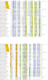
Multiple alignment of JAB domain containing proteins. Coloring is indicative of 80% consensus. The coloring scheme, consensus abbreviations and secondary structure representations are as described in the legend to Figure 2. The secondary structure, shown on the first line of the alignment, is derived from a JAB crystal structure whose primary sequence is found on the second line of the alignment, with PDB identifier shaded in gold. Conserved histidine and acidic residues (ED) are colored yellow and shaded in red. The conserved active site serine residue is colored light gray and shaded in teal. The conserved cysteine found in a subset of JABs (marked with an asterisk) are shaded blue and colored white. The alignment is grouped according to families, with family names listed to the right. Also provided are references to the appropriate row on Table 1, which describes a particular JAB containing operon.
Figure 6
Figure 6
Multiple alignment of E2 (UBC)-like proteins with a special emphasis on bacterial versions. PDB identifiers of primary sequences derived from crystal structures are shaded in gold. Coloring is indicative of 55% consensus. The secondary structure, shown on the second line of the alignment, is derived from a general consensus of the secondary structure features from the different crystal structures shown in the alignment. Other features of the alignment are the same as in Figure 2, including coloring scheme, consensus abbreviations and secondary structure representations. Additionally, conserved polar residues (p; CDEHKNQRST) are colored blue. The strongly conserved proline and asparagine residues are colored purple brown respectively. The strongly conserved cysteine and histidine residues described in the text are shaded red and are also marked with an asterisk above their positions in the alignment. The major families of bacterial E2s are shown to the right. Also shown are the row numbers in Table 1, where a particular family is described. See the legend to Figure 2 for species abbreviations.
Figure 7
Figure 7
Network diagram of ThiS/MoaD-like β-grasp domains. The interaction network depicted here represents the known functional associations (arrows colored orange), the associations suggested by domain architectures (arrows colored green), and the associations suggested by gene neighborhood (arrows colored gray) between pairs of domains, as described in the text. The directionality of the network interactions, as indicated by an arrowhead, represents the order of a domain pair from the amino- to the carboxyl-terminus of the domain architecture or from the 5' to 3' end of a gene neighborhood. Lines with arrowheads at both ends represent domain pairs found both amino-terminal and carboxyl-terminal to each other in domain architectures or 5' to 3' in operonic contexts. The primary 'hubs' of the network are highlighted prominently. Domains are not exactly to scale. Selected interactions are encircled by small ellipses connected to the labels describing the functional role of the interaction. The labels are portrayed as large black ellipses with white lettering. MBL, metallo-β-lactamase domain; OAHS hyd, O-acetylhomoserine sulfhydrylase; PDOR, pyridine disulfide oxidoreductase; Rhod, Rhodanese-like domain; Toluene mono, toluene mono-oxygenase; ZnR, zinc-ribbon containing domain.

Similar articles

Cited by

References

    1. Alberts B, Johnson A, Lewis J, Raff M, Roberts K, Walter P. Molecular Biology of the Cell, (book and CD-ROM) 4. New York, NY: Garland Science Publishing; 2002.
    1. Hershko A, Ciechanover A. The ubiquitin system. Annu Rev Biochem. 1998;67:425–479. doi: 10.1146/annurev.biochem.67.1.425. - DOI - PubMed
    1. Ciechanover A, Orian A, Schwartz AL. Ubiquitin-mediated proteolysis: biological regulation via destruction. Bioessays . 2000;22:442–451. doi: 10.1002/(SICI)1521-1878(200005)22:5<442::AID-BIES6>3.0.CO;2-Q. - DOI - PubMed
    1. Ardley HC, Robinson PA. E3 ubiquitin ligases. Essays Biochem. 2005;41:15–30. - PubMed
    1. Wertz IE, O'Rourke KM, Zhou H, Eby M, Aravind L, Seshagiri S, Wu P, Wiesmann C, Baker R, Boone DL, et al. De-ubiquitination and ubiquitin ligase domains of A20 downregulate NF-kappaB signalling. Nature. 2004;430:694–699. doi: 10.1038/nature02794. - DOI - PubMed

Publication types

LinkOut - more resources