. 2006;8(4):R42.
doi: 10.1186/bcr1524.

Beta3 integrin and Src facilitate transforming growth factor-beta mediated induction of epithelial-mesenchymal transition in mammary epithelial cells

Affiliations

Beta3 integrin and Src facilitate transforming growth factor-beta mediated induction of epithelial-mesenchymal transition in mammary epithelial cells

Amy J Galliher et al. Breast Cancer Res. 2006.

Abstract

Introduction: Transforming growth factor (TGF)-beta suppresses breast cancer formation by preventing cell cycle progression in mammary epithelial cells (MECs). During the course of mammary tumorigenesis, genetic and epigenetic changes negate the cytostatic actions of TGF-beta, thus enabling TGF-beta to promote the acquisition and development of metastatic phenotypes. The molecular mechanisms underlying this conversion of TGF-beta function remain poorly understood but may involve signaling inputs from integrins.

Methods: beta3 Integrin expression or function in MECs was manipulated by retroviral transduction of active or inactive beta3 integrins, or by transient transfection of small interfering RNA (siRNA) against beta3 integrin. Altered proliferation, invasion, and epithelial-mesenchymal transition (EMT) stimulated by TGF-beta in control and beta3 integrin manipulated MECs was determined. Src involvement in beta3 integrin mediated alterations in TGF-beta signaling was assessed by performing Src protein kinase assays, and by interdicting Src function pharmacologically and genetically.

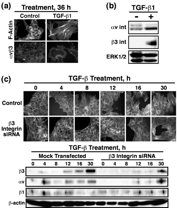
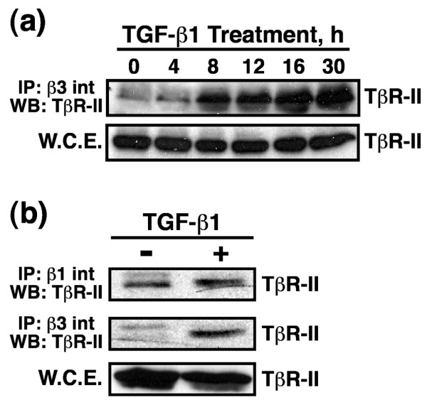
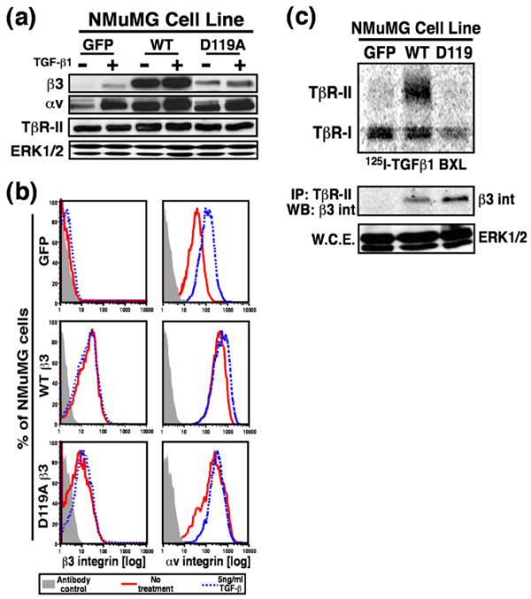
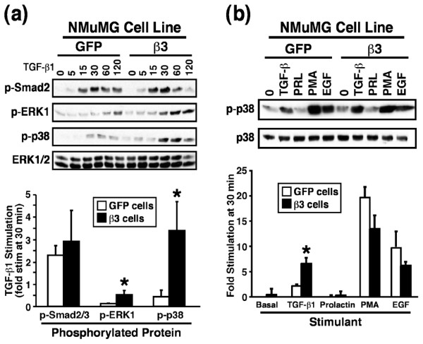
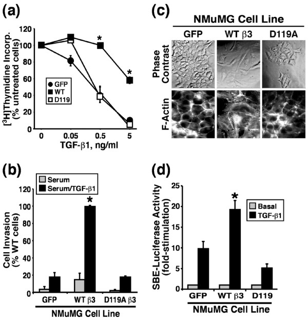
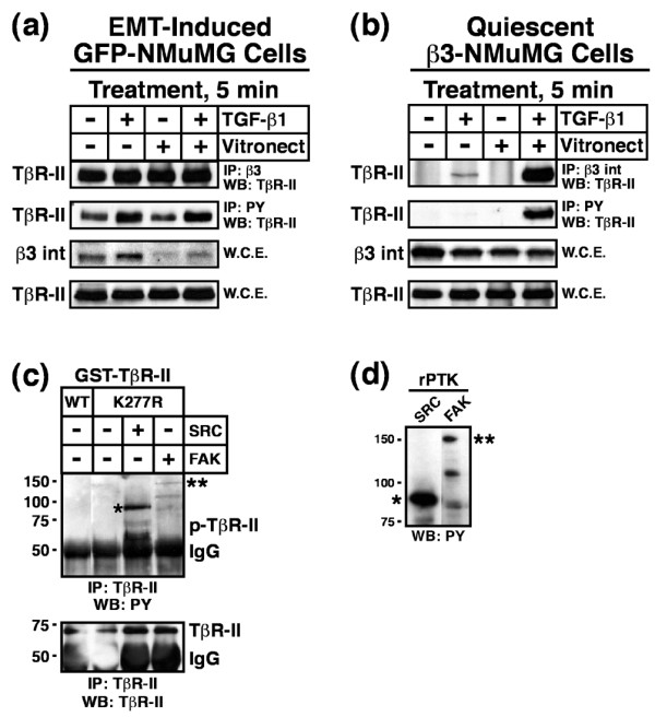
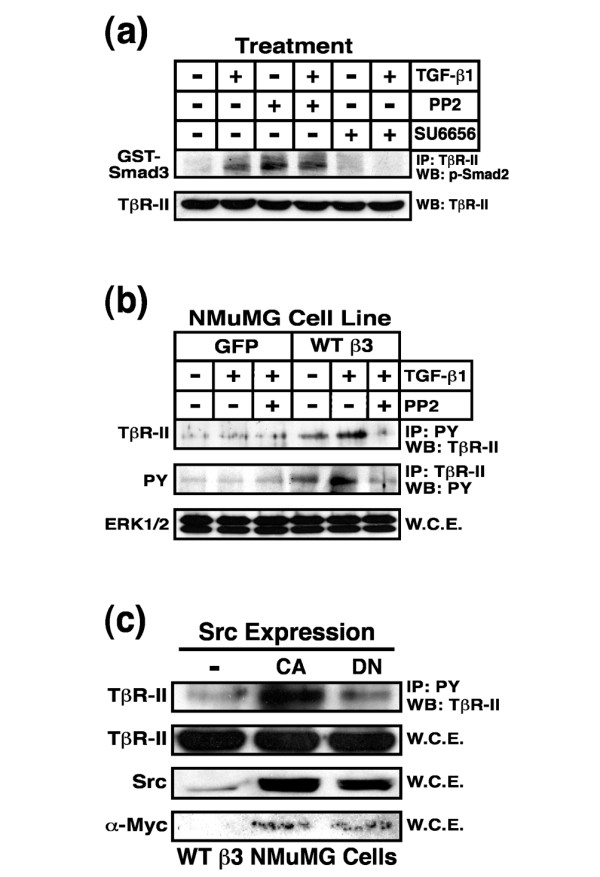
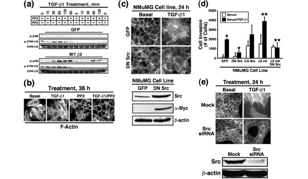
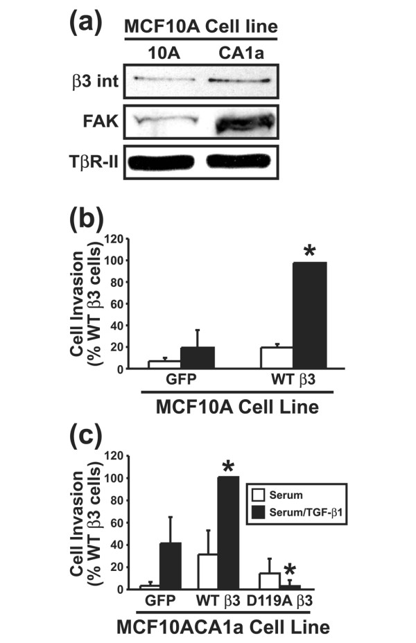
Results: TGF-beta stimulation induced alphavbeta3 integrin expression in a manner that coincided with EMT in MECs. Introduction of siRNA against beta3 integrin blocked its induction by TGF-beta and prevented TGF-beta stimulation of EMT in MECs. beta3 integrin interacted physically with the TGF-beta receptor (TbetaR) type II, thereby enhancing TGF-beta stimulation of mitogen-activated protein kinases (MAPKs), and of Smad2/3-mediated gene transcription in MECs. Formation of beta3 integrin:TbetaR-II complexes blocked TGF-beta mediated growth arrest and increased TGF-beta mediated invasion and EMT. Dual beta3 integrin:TbetaR-II activation induced tyrosine phosphorylation of TbetaR-II, a phosphotransferase reaction mediated by Src in vitro. Inhibiting Src activity in MECs prevented the ability of beta3 integrin to induce TbetaR-II tyrosine phosphorylation, MAPK activation, and EMT stimulated by TGF-beta. Lastly, wild-type and D119A beta3 integrin expression enhanced and abolished, respectively, TGF-beta stimulation of invasion in human breast cancer cells.

Conclusion: We show that beta3 integrin alters TGF-beta signaling in MECs via Src-mediated TbetaR-II tyrosine phosphorylation, which significantly enhanced the ability of TGF-beta to induce EMT and invasion. Our findings suggest that beta3 integrin interdiction strategies may represent an innovative approach to re-establishing TGF-beta mediated tumor suppression in progressing human breast cancers.


Figures

Figure 1
Figure 1
TGF-β1 mediated EMT increases cell surface αvβ3 integrin expression in NMuMG cells. NMuMG cells were incubated in the absence or presence of TGF-β1 (5 ng/ml) for 0–36 hours and assayed for EMT. (a) Alterations in actin cytoskeletal architecture were visualized by direct rhodamine-phalloidin immunofluorescence and αvβ3 integrin by indirect immunofluorescence using anti-αvβ3 integrin (LM609) antibodies. Shown are representative images from a single experiment that was repeated twice with identical results. (b) Detergent-solubilized whole cell extracts (50 μg/lane) were prepared to analyze expression of the αv and β3 integrin subunits by immunoblotting. Differences in protein loading were monitored by reprobing striped blots with anti-ERK1/2 antibodies because, unlike the increase in β-actin expression observed (data not shown) ERK1/2 expression was unaltered in NMuMG cells in response to TGF-β1 treatment. (c) β3 Integrin deficient NMuMG cells were generated by siRNA transfection. The effects of β3 integrin deficiency on TGF-β mediated EMT was visualized by direct FITC-phalloidin immunofluorescence. Shown are representative images from a single experiment that was repeated twice with identical results. Detergent-solubilized whole cell extracts (50 μg/lane) were prepared to analyze expression of the αv, β3, and β1 integrin subunits by immunoblotting. Differences in protein loading were monitored by reprobing striped blots with anti-β-actin antibodies. EMT, epithelial-mesenchymal transitions; ERK, extracellular signal-regulated kinase; siRNA, small interfering RNA; TGF, transforming growth factor.
Figure 2
Figure 2
TGF-β1 mediated EMT induces β3 integrin:TβR-II complex formation on the cell surface of NMuMG cells. NMuMG cells were incubated in the absence or presence of TGF-β1 (5 ng/ml) for 0–30 hours to induce EMT. Detergent-solubilized whole cell extracts (1 mg/tube) were immunoprecipitated with (a,b) anti-β3 or (b) anti-β1 integrin antibodies, and subsequently immunoblotted with anti-TβR-II antibodies. Shown are representative immunoblots from a single experiment that was repeated twice with identical results. EMT, epithelial-mesenchymal transitions; TβR; TGF receptor; TGF, transforming growth factor; WCE, whole cell extracts.
Figure 3
Figure 3
β3 Integrin overexpression in NMuMG cells. NMuMG cells were engineered by bi-cistronic retroviral transduction to stably express GFP alone (control), WT β3 integrin (WT) and GFP, or its inactive mutant D119A and GFP. (a) Detergent-solubilized whole cell extracts (50 μg/lane) were prepared to analyze expression of the αv and β3 integrin subunits, and of TβR-II. Differences in protein loading were monitored by immunoblotting ERK1/2, whose expression in NMuMG cells was not altered by TGF-β treatment. (b) The percentage of NMuMG cells expressing human recombinant β3 integrin or endogenous αv integrin was determined by FACS analysis of cells incubated with either anti-human β3 integrin or anti-mouse αv integrin antibodies. Histograms depict β3 and αv expression only in GFP-positive NMuMG cells and are from a representative experiment that was repeated twice with similar results. (c) Iodinated TGF-β1 was bound and chemically cross-linked to control (GFP), WT, and D119A β3 integrin expressing cells. Cytokine:receptor complexes were isolated by immunoprecipitation with anti-TβR-II antibodies, fractionated through 7.5% SDS-PAGE gels, and immobilized electrophoretically to nitrocellulose membranes. The upper panel shows a representative phosphor image of iodinated TGF-β1 bound to TβR-I and TβR-II. The middle panel shows that β3 integrin was present in isolated iodinated TGF-β1:receptor complexes as detected by anti-β3 integrin Western blot analysis of anti-TβR-II immunocomplexes. Differences in protein loading were monitored by probing detergent-solubilized whole cell extracts (50 μg/lane) with anti-ERK1/2 antibodies (bottom panel). BXL, binding and cross-linking; ERK, extracellular signal-regulated kinase; GFP, green fluorescent protein; WT, wild type; TβR, TGF receptor; TGF, transforming growth factor; WCE, whole cell extracts.
Figure 4
Figure 4
β3 Integrin enhances MAPK activation by TGF-β in NMuMG cells. (a) Control or β3 integrin expressing NMuMG cells were serum starved for 4 hours prior to TGF-β1 stimulation (5 ng/ml) for 0–120 min. Afterward, the activation status of Smad2, ERK1/2, and p38 MAPK was determined by immunoblot analysis using phospho-specific antibodies. Reprobing stripped membranes with anti-ERK1/2 antibodies monitored differences in protein loading. (b) Control or β3 integrin-expressing NMuMG cells were serum starved for 4 hours before stimulation with TGF-β1 (5 ng/ml), prolactin (100 ng/ml), PMA (10 ng/ml), or EGF (100 ng/ml) for 30 min. Afterward, the activation status of p38 MAPK was determined by immunoblot analysis using phospho-specific antibodies. Reprobing stripped membranes with anti-pan p38 MAPK antibodies was used to monitor differences in protein loading. Accompanying graphs depict the mean (± standard error) fold increase of p38 MAPK stimulation observed in three independent experiments (*P < 0.05). EGF, epidermal growth factor; ERK, extracellular signal-regulated kinase; MAPK, mitogen-activated protein kinase; PMA, 4α-phorbol 12-myristate 13-acetate; TGF, transforming growth factor.
Figure 5
Figure 5
β3 Integrin expression blocks TGF-β stimulated growth arrest but enhances TGF-β stimulated invasion and EMT in NMuMG cells. (a) Control (GFP), WT, or D119A β3 integrin expressing NMuMG cells were stimulated with increasing concentrations of TGF-β1 for 48 hours. Cellular DNA was radiolabeled with [3H]thymidine and quantified by scintillation counting. Data are the means (± standard error) of three independent experiments presented as the percentage of [3H]thymidine incorporation normalized to untreated cells. (b) Control (GFP) and β3 integrin expressing NMuMG cells were allowed to invade through Matrigel matrices in the absence or presence of TGF-β1 (5 ng/ml) for 36 hours. Values are the mean (± standard error) of three independent experiments presented as the percentage invasion relative to TGF-β stimulated β3 integrin expressing NMuMG cells. (c) The cell morphology of control (GFP), WT, and D119A β3 integrin expressing NMuMG cells was monitored by phase contrast microscopy, and alterations in actin cytoskeletal architecture was visualized by direct rhodamine-phalloidin immunofluorescence. Shown are representative images from a single experiment that was repeated twice with identical results. (d) Control (GFP), WT, or D119A β3 integrin expressing NMuMG cells were transiently transfected with pSBE-luciferase and pCMV-β-gal cDNAs, and subsequently were stimulated with TGF-β1 (5 ng/ml) for 24 hours. Afterward, luciferase and β-gal activities contained in detergent-solubilized cell extracts were measured. Values are the mean (± standard error) luciferase activities observed in three independent experiments normalized to maximal reporter gene expression measured in unstimulated cells. EMT, epithelial-mesenchymal transitions; GFP, green fluorescent protein; TGF, transforming growth factor; WT, wild type; SBE, Smad binding element.
Figure 6
Figure 6
Dual receptor activation induces Src mediated TβR-II tyrosine phosphorylation. (a) GFP-expressing NMuMG cells were stimulated with TGF-β1 (5 ng/ml) for 36 hours to induce β3 integrin expression and EMT. Afterward, the cells were dissociated and re-plated either onto plastic or vitronectin-coated wells in the absence or presence of TGF-β1 (5 ng/ml) for 5 min. Afterward, detergent-solubilized whole cell extracts were prepared (1 mg/tube) and subsequently immunoprecipitated with anti-phosphotyrosine or anti-β3 integrin antibodies as indicated. The presence of TβR-II in precipitated immunocomplexes was determined by Western blotting with anti-TβR-II antibodies. Differences in protein loading were monitored by immunoblotting whole cell extracts (50 mg/lane) for β3 integrin and TβR-II. Shown are representative immunoblots from a single experiment that was repeated twice with similar results. (b) Resting WT β3 integrin-expressing NMuMG cells were dissociated and re-plated as above. Afterward, TβR-II tyrosine phosphorylation and complex formation with β3 integrin was determined by immunoblotting as above. Shown are representative immunoblots from a single experiment that was repeated three times with identical results. (c) Active recombinant Src (1 unit/reaction) or FAK (0.2 μg/reaction) kinases were added to protein kinase reaction mixtures containing 1 μg/tube of either kinase active (WT) or inactive (K277R) GST-TβR-II. Phosphorylation reactions were stopped after 30 min and the phosphorylation status of TβR-II was determined by immunoprecipitation of diluted protein kinase reaction mixtures with anti-TβR-II antibodies. Afterward, immobilized immunocomplexes were probed sequentially with anti-phosphotyrosine antibodies, followed by anti-TβR-II antibodies as indicated (*autophosphorylated recombinant Src; **autophosphorylated recombinant FAK). Data are from a representative experiment that was repeated three times with similar results. (d) Recombinant Src and FAK were incubated in protein kinase assay buffer for 30 min at 30°C. The data show the autophosphorylation on tyrosine residues of recombinant Src (*) and FAK (**) as determined by immunoblotting with anti-phosphotyrosine antibodies. Data are from a representative experiment that was repeated at least three times with similar results. EMT, epithelial-mesenchymal transitions; FAK, focal adhesion kinase; GST, glutathione S-transferase; TβR, TGF-β receptor; TGF, transforming growth factor; WCE, whole cell extracts; WT, wild type.
Figure 7
Figure 7
Src inhibition blocks TβR-II tyrosine phosphorylation in NMuMG cells. (a) NMuMG cells were pretreated for 60 min with either PP2 (10 μmol/l) or SU6656 (10 μmol/l). Afterward, the cells were stimulated with TGF-β1 (5 ng/ml) for 20 min, and subsequently were lysed and immunoprecipitated (1 mg/tube) with anti-TβR-II antibodies. The resulting immunocomplexes were used to phosphorylate recombinant GST-Smad3, which was visualized by immunoblotting with anti-phospho-Smad2 antibodies. Data are from a representative experiment that was repeated three times with similar results. (b) Control or β3 integrin expressing NMuMG cells were stimulated with TGF-β1 (5 ng/ml) in the absence or presence the Src inhibitor PP2 (10 μmol/l), as indicated. Afterward, detergent-solubilized whole cell extracts were prepared, immunoprecipitated, and subjected to Western blot analysis either with anti-phosphotyrosine (2 mg WCE/tube) or anti-TβR-II (1 mg WCE/tube) antibodies as indicated. Differences in protein loading were monitored by probing detergent-solubilized whole cell extracts (50 μg/lane) with anti-ERK1/2 antibodies (bottom panel). (c) WT β3 integrin expressing NMuMG cells were transiently transfected with cDNAs encoding either constitutively active (CA) or dominant negative (DN) Src. Afterward, detergent solubilized whole cell extracts were prepared, immunoprecipitated with anti-phosphotyrosine (1 mg WCE/tube), and subjected to WB analysis with anti-TβR-II antibodies. Total Src expression was monitored by probing detergent solubilized whole cell extracts (50 μg/lane) with anti-Src antibodies, whereas exogenous Src expression was confirmed by reprobing stripped membranes with anti-Myc antibodies. Differences in protein loading were monitored by probing stripped membranes with anti-TβR-II antibodies. ERK, extracellular signal-regulated kinase; GST, glutathione S-transferase; IP, immunoprecipitated; PY, phosphotyrosine; TβR, TGF-β receptor; TGF, transforming growth factor; WB, Western blot; WCE, whole cell extracts; WT, wild type.
Figure 8
Figure 8
Src inhibition blocks β3 integrin and TGF-β mediated MAPK activation, EMT, and invasion in NMuMG cells. (a) Control or β3 integrin expressing NMuMG cells were serum starved for 4 hours before TGF-β1 (5 ng/ml) stimulation in the absence or presence of either PP2 (10 μmol/l) or its inactive counterpart PP3 (10 μmol/l). Afterward, the activation status of ERK1/2 and p38 MAPK was determined by immunoblot analysis using phospho-specific antibodies. Re-probing stripped membranes with anti-ERK1/2 antibodies monitored differences in protein loading. Data are from a representative experiment that was repeated twice with similar results. (b) β3 Integrin expressing NMuMG cells were stimulated with TGF-β1 (5 ng/ml) for 36 hours in the absence or presence of PP2 (10 μmol/l). Afterward, alterations in actin cytoskeletal architecture were visualized by direct rhodamine-phalloidin immunofluorescence. Shown are representative images from a single experiment that was repeated twice with identical results. (c) Control (GFP) or DN Src-expressing NMuMG cells were stimulated with TGF-β1 (5 ng/ml) for 24 hours. Afterward, alterations in actin cytoskeletal architecture were visualized by direct rhodamine-phalloidin immunofluorescence. Total Src expression was monitored by probing detergent-solubilized whole cell extracts (50 μg/lane) with anti-Src antibodies, whereas exogenous Src expression was visualized using anti-Myc antibodies. Immunoblotting stripped membranes with anti-β-actin antibodies served to monitor differences in protein loading. Shown are representative images from a single experiment that was repeated twice with identical results. (d) Control (GFP), DN Src, CA Src, β3 integrin, and β3 integrin/DN Src expressing NMuMG cells were allowed to invade through Matrigel matrices in the absence or presence of TGF-β1 (5 ng/ml) for 36 hours. Values are the mean (± standard error) of three independent experiments presented as the number of cells invaded per well. (e) Src-deficient NMuMG cells were generated by siRNA transfection. The effects of Src deficiency on TGF-β mediated EMT were visualized by direct FITC-phalloidin immunofluorescence. Shown are representative images from a single experiment that was repeated twice with identical results. Detergent-solubilized whole cell extracts (50 μg/lane) were prepared to monitor Src expression by immunoblotting with anti-Src antibodies. Differences in protein loading were monitored by re-probing striped blots with anti-β-actin antibodies. CA, constitutively active; DN, dominant negative; ERK, extracellular signal-regulated kinase; GFP, green fluorescent protein; MAPK, mitogen-activated protein kinase; TGF, transforming growth factor; WB, Western blot; WT, wild type.
Figure 9
Figure 9
β3 Integrin expression increases in malignant human MECs and regulates invasion stimulated by TGF-β. (a) Detergent-solubilized whole cell extracts (100 μg/lane) of MCF10A and MCF10CA1a cells were prepared to analyze expression of the β3 integrin subunit and FAK by immunoblotting. Differences in protein loading were monitored by probing striped blots with anti-TβR-II antibodies. Control (GFP) and β3 integrin expressing (b) MCF10A cells or (c) MCF10CA1a cells were allowed to invade through Matrigel matrices in the absence or presence of TGF-β1 (5 ng/ml) for 36 hours. Values are the mean (± standard error) of three independent experiments presented as the percentage invasion relative to TGF-β stimulated WT β3 integrin expressing cells. FAK, focal adhesion kinase; MEC, mammary epithelial cell; TβR, TGF-β receptor; TGF, transforming growth factor; WT, wild type.

Similar articles

Cited by

References

    1. Shi Y, Massague J. Mechanisms of TGF-β signaling from cell membrane to the nucleus. Cell. 2003;113:685–700. doi: 10.1016/S0092-8674(03)00432-X. - DOI - PubMed
    1. Bakin AV, Rinehart C, Tomlinson AK, Arteaga CL. p38 mitogen-activated protein kinase is required for TGFβ-mediated fibroblastic transdifferentiation and cell migration. J Cell Sci. 2002;115:3193–3206. - PubMed
    1. Bakin AV, Tomlinson AK, Bhowmick NA, Moses HL, Arteaga CL. Phosphatidylinositol 3-kinase function is required for transforming growth factor b-mediated epithelial to mesenchymal transition and cell migration. J Biol Chem. 2000;275:36803–36810. doi: 10.1074/jbc.M005912200. - DOI - PubMed
    1. Perlman R, Schiemann WP, Brooks MW, Lodish HF, Weinberg RA. TGF-β-induced apoptosis is mediated by the adapter protein Daxx that facilitates JNK activation. Nat Cell Biol. 2001;3:708–714. doi: 10.1038/35087019. - DOI - PubMed
    1. Zavadil J, Bitzer M, Liang D, Yang YC, Massimi A, Kneitz S, Piek E, Bottinger EP. Genetic programs of epithelial cell plasticity directed by transforming growth factor-β. Proc Natl Acad Sci USA. 2001;98:6686–6691. doi: 10.1073/pnas.111614398. - DOI - PMC - PubMed

LinkOut - more resources