. 2006;8(4):R124.
doi: 10.1186/ar2013.

Expression profiling of metalloproteinases and their inhibitors in synovium and cartilage

Affiliations

Expression profiling of metalloproteinases and their inhibitors in synovium and cartilage

Rose K Davidson et al. Arthritis Res Ther. 2006.

Abstract

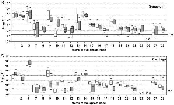
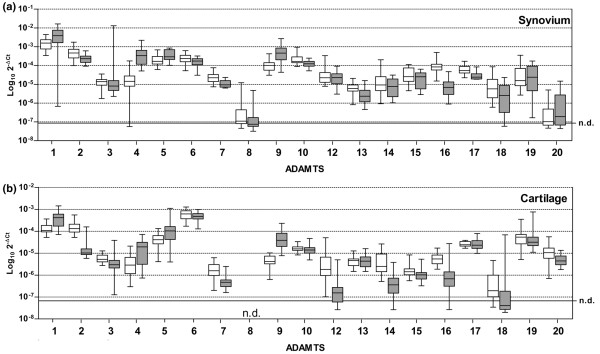
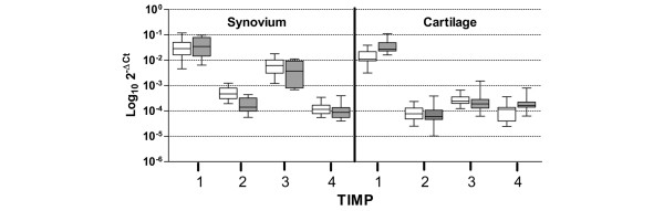
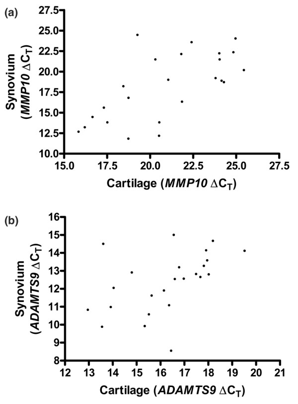
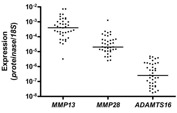
Cartilage destruction in osteoarthritis (OA) is thought to be mediated by two main enzyme families; the matrix metalloproteinases (MMPs) are responsible for cartilage collagen breakdown, whereas enzymes from the 'a disintegrin and metalloproteinase domain with thrombospondin motifs' (ADAMTS) family mediate cartilage aggrecan loss. Tissue inhibitors of metalloproteinases (TIMPs) regulate the activity of these enzymes. Although cartilage destruction in OA might be driven by the chondrocyte, low-grade synovitis is reported in patients with all grades of this disease. Our earlier work profiling these gene families in cartilage identified a number of genes that are regulated in OA, which are hence implicated in the disease process. Because the synovium might contribute to cartilage-matrix destruction in OA, we have extended the screening in the current study. We have profiled MMP, ADAMTS and TIMP genes in both cartilage and synovium from patients with either OA of the hip or a fracture to the neck of femur (NOF), giving a more complete picture of proteolysis in this disease. The four most significantly upregulated genes (P < 0.0001) in OA synovium compared to the fractured NOF are MMP28, ADAMTS16, ADAMTS17 and TIMP2. For MMP9, MMP10, MMP12, MMP17, MMP23, MMP28, ADAMTS4, and ADAMTS9, there is a significant correlation between expression levels in the synovium and cartilage, suggesting similar mechanisms of regulation. Additionally, we have shown that in cartilage the median level of steady-state mRNA for MMP13 is approximately 20-fold higher than MMP28 and approximately 1,500-fold higher than ADAMTS16, with expression of this latter gene approximately 150-fold higher in synovium than cartilage. This study is the most comprehensive analysis of the metzincin family of proteinases in the joint to date and has identified several proteinase genes not previously reported to be expressed or regulated in synovium.


Figures

Figure 1
Figure 1
Comparative expression of MMP genes in the fractured NOF (filled boxes) compared with OA (open boxes) in hip (a) synovium and (b) cartilage. The expression level of each gene was determined, as described in Materials and methods, and normalized to the level of 18S rRNA gene expression using ΔCT [CT(target gene) – CT(18S)]. The box-and-whisker plot shows median values, and each section represents a quartile of the data. Samples on or below the dotted line have CT(target gene) = 40 and are therefore not detectable (n.d.).
Figure 2
Figure 2
Comparative expression of ADAMTS genes in the fractured NOF (filled boxes) compared with OA (open boxes) hip (a) synovium and (b) cartilage. The expression level of each gene was determined, as described in Materials and methods, and normalized to the level of 18S rRNA gene expression using ΔCT [CT(target gene) – CT(18S)]. The box-and-whisker plot shows median values, and each section represents a quartile of the data. Samples on or below the dotted line have CT(target gene) = 40 and are therefore not detectable (n.d.).
Figure 3
Figure 3
Comparative expression of TIMP genes in the fractured NOF (filled boxes) compared with OA (open boxes) hip synovium and cartilage. The expression level of each gene was determined, as described in Materials and methods, and normalized to the level of 18S rRNA gene expression using ΔCT [CT(target gene) – CT(18S)]. The box-and-whisker plot shows median values, and each section represents a quartile of the data.
Figure 4
Figure 4
Correlation analysis of gene expression in cartilage compared with synovium. Correlation plots for (a) MMP10 and (b) ADAMTS9 expression in cartilage and synovium taken from the same joint. Correlation coefficients are as follows: MMP10, r = 0.645 (P = 0.0006); and ADAMTS9, r = 0.590 (P = 0.0024).
Figure 5
Figure 5
Comparison of MMP13, MMP28 and ADAMTS16 expression levels in cartilage. Expression of each gene was quantified against standard curves constructed using purified cDNA and normalized to 18S expression, which was similarly quantified. Median values are marked.

References

    1. The Arthritis Research Campaign, UK http://www.arc.org.uk/about_arth/bigpic.htm
    1. Wieland HA, Michaelis M, Kirschbaum BJ, Rudolphi KA. Osteoarthritis – an untreatable disease? Nat Rev Drug Discov. 2005;4:331–344. doi: 10.1038/nrd1693. - DOI - PubMed
    1. Roughley PJ. Articular cartilage and changes in arthritis: noncollagenous proteins and proteoglycans in the extracellular matrix of cartilage. Arthritis Res. 2001;3:342–347. doi: 10.1186/ar326. - DOI - PMC - PubMed
    1. Eyre D. Collagen of articular cartilage. Arthritis Res. 2002;4:30–35. doi: 10.1186/ar380. - DOI - PMC - PubMed
    1. Murphy G, Knauper V, Atkinson S, Butler G, English W, Hutton M, Stracke J, Clark I. Matrix metalloproteinases in arthritic disease. Arthritis Res. 2002;4(Suppl 3):S39–49. doi: 10.1186/ar572. - DOI - PMC - PubMed

Publication types

MeSH terms