Comparative Study
. 2006;8(4):R115.
doi: 10.1186/ar2002. Epub 2006 Jul 19.

A model of anti-angiogenesis: differential transcriptosome profiling of microvascular endothelial cells from diffuse systemic sclerosis patients

Affiliations
Comparative Study

A model of anti-angiogenesis: differential transcriptosome profiling of microvascular endothelial cells from diffuse systemic sclerosis patients

Betti Giusti et al. Arthritis Res Ther. 2006.

Expression of concern in

Abstract

The objective of this work was to identify genes involved in impaired angiogenesis by comparing the transcriptosomes of microvascular endothelial cells from normal subjects and patients affected by systemic sclerosis (SSc), as a unique human model disease characterized by insufficient angiogenesis. Total RNAs, prepared from skin endothelial cells of clinically healthy subjects and SSc patients affected by the diffuse form of the disease, were pooled, labeled with fluorochromes, and hybridized to 14,000 70 mer oligonucleotide microarrays. Genes were analyzed based on gene expression levels and categorized into different functional groups based on the description of the Gene Ontology (GO) consortium to identify statistically significant terms. Quantitative PCR was used to validate the array results. After data processing and application of the filtering criteria, the analyzable features numbered 6,724. About 3% of analyzable transcripts (199) were differentially expressed, 141 more abundantly and 58 less abundantly in SSc endothelial cells. Surprisingly, SSc endothelial cells over-express pro-angiogenic transcripts, but also show up-regulation of genes exerting a powerful negative control, and down-regulation of genes critical to cell migration and extracellular matrix-cytoskeleton coupling, all alterations that provide an impediment to correct angiogenesis. We also identified transcripts controlling haemostasis, inflammation, stimulus transduction, transcription, protein synthesis, and genome organization. An up-regulation of transcripts related to protein degradation and ubiquitination was observed in SSc endothelial cells. We have validated data on the main anti-angiogenesis-related genes by RT-PCR, western blotting, in vitro angiogenesis and immunohistochemistry. These observations indicate that microvascular endothelial cells of patients with SSc show abnormalities in a variety of genes that are able to account for defective angiogenesis.


Figures
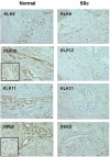
Figure 1
Figure 1
Immunohistochemical evidence of differential expression of tissue kallikrein (KLK)9, KLK11, KLK12, and DSG2 in microvascular endothelial cells (MVECs) from normal subjects and patients with systemic sclerosis (SSc). Each panel is representative of the pattern of immune staining with each antibody obtained for the six normal and six SSc patients subjected to skin biopsies from which MVECs were prepared. The inset in the micrograph of KLK12 shows negative staining using isotype control rabbit IgG (the isotype used for KLK9 and KLK12), while that shown in the micrograph of DSG shows negative staining for mouse IgG (the isotype used for KLK11 and DSG). Original magnification, 200×.
Figure 2
Figure 2
Expression and role of plexin B1 (PLXNB1) in endothelial cell invasion and capillary morphogenesis. (a) Western blotting of 60 μg protein from cell lysates of microvascular endothelial cells (MVECs) from normal subjects (N-MVECs) and patients with systemic sclerosis (SSc-MVECs) with anti-PLXNB1 antibodies. Each lane represents western blotting of MVECs obtained from a single patient. Actin was used as an internal reference standard. Numbers on the right represent the molecular weight expressed in kDa. (b) Effect of anti-PLXNB1 antibodies (3 μg/ml) on matrigel invasion of N-MVECs. The effect of irrelevant rabbit IgG is also shown. Numbers on the x-axis refer to the total number of cells migrated through the matrigel after 6 hours. Data are the mean ± standard deviation of three experiments performed in triplicate in three N-MVEC lines. The asterisk indicates that values are significantly different from the values of control (p < 0.05). (c) Effect of anti-PLXNB1 antibodies on capillary morphogenesis of N-MVECs. N-MVECs were plated on Matrigel (60 × 103/ml), in complete MCDB medium, supplemented with 30% fetal calf serum, and 20 μg/ml endothelial cells growth supplement. N-MVEC spontaneously form anastomosing cords of cells resembling a capillary plexus, which are well organized by 6 hours. The process of endothelial cell organization after 6 hours is impaired in the presence of 3 μg/ml of anti-PLXNB1 rabbit polyclonal antibodies. Irrelevant rabbit IgG gave results similar to control untreated N-MVECs (not shown). These data are representative of three different experiments performed on three N-MVEC cell lines (100× magnification). Numbers reported within each panel indicate the percent of the photographic field occupied by cells ± standard deviation. The asterisk indicates that values are significantly different from the values of control N-MVECs at 6 hours after plating on Matrigel (p < 0.05).
Figure 3
Figure 3
Expression and role of pent(r)axin 3 (PTX3) in endothelial cell invasion and capillary morphogenesis. (a) Western blotting of 60 μg protein from cell lysates of microvascular endothelial cells (MVECs) from normal subjects (N-MVECs) and patients with systemic sclerosis (SSc-MVECs) with anti-PTX3 antibodies. Each lane represents western blotting of MVECs obtained from a single patient. Actin was used as an internal reference standard. Numbers on the right represent the molecular weight expressed in kDa. (b) Effect of PTX3 antibodies (3 μg/ml) on matrigel invasion of N-MVECs. The effect of irrelevant rat IgG is also shown. Numbers on the x-axis refer to the total number of cells migrated through the matrigel after 6 hours. The presence of anti-PTX3 antibodies is able to revert inhibition of matrigel invasion induced by SSc-MVECs conditioned medium (C.M.). Data are the mean ± standard deviation of three experiments performed in triplicate in three N-MVEC lines. An asterisk indicates that values are significantly different from the values of control (p < 0.05). (c) Effect of anti-PTX3 antibodies on capillary morphogenesis of N-MVECs. Experimental conditions were as described in the legend to Figure 1. Conditioned medium from SSc-MVECs was able to impair capillary morphogenesis observed after 6 hours from plating (panel c). The process of endothelial cell organization after 6 hours was partially restored in the presence of 3 μg/ml of anti-PTX3 rat monoclonal antibodies (panel d), which showed only a small effect when added to control N-MVECs (panel b). Irrelevant rat IgG did not show any effect (not shown). These data are representative of three different experiments performed on three N-MVEC cell lines (100× magnification). Numbers reported within each panel indicate the percent of the photographic field occupied by cells ± standard deviation. An asterisk indicates that values are significantly different from the values of control N-MVECs at 6 hours after plating on Matrigel (p < 0.05).
Figure 4
Figure 4
Expression and role of desmoglein (DSG)2 in endothelial cell invasion and capillary morphogenesis. (a) Western blotting of 60 μg protein from cell lysates of microvascular endothelial cells (MVECs) from normal subjects (N-MVECs) and patients with systemic sclerosis (SSc-MVECs) with anti-DSG2 antibodies. Each lane represents western blotting of MVECs obtained from a single patient. Actin was used as an internal reference standard. Numbers on the right represent the molecular weight expressed in kDa. (b) Effect of anti-DSG2 antibodies (3 μg/ml) on matrigel invasion of N-MVECs. The effect of irrelevant mouse IgG is also shown. Numbers on the x-axis refer to the total number of cells migrated through the matrigel after 6 hours. Data are the mean ± standard deviation of three experiments performed in triplicate in three N-MVEC lines. The asterisk indicates that values are significantly different from the values of control (p < 0.05). (c) Effect of anti-DSG2 antibodies on capillary morphogenesis of N-MVECs. See the legend to Figure 1 for experimental details. Irrelevant mouse IgG gave results similar to control untreated N-MVECs (not shown). These data are representative of three different experiments performed on three N-MVEC cell lines (100× magnification). Numbers reported within each panel indicate the percent of the photographic field occupied by cells ± standard deviation. The asterisk indicates that values are significantly different from the values of control N-MVECs at 6 hours after plating on Matrigel (p < 0.05).
Figure 5
Figure 5
Impairment of angiogenesis in systemic sclerosis. Genes differentially expressed in microvascular endothelial cells (MVECs) from patients with systemic sclerosis (SSc-MVECs) are grouped according to Table 3. Upward and downward arrows indicate up-regulation and down-regulation, respectively, in SSc-MVECs. Differentially expressed genes that we propose to be critical to the altered angiogenic process in SSc are reported in italics in each sub-group. In SSc, the powerful angiogenic instruction of Vascular Endothelial Growth Factor (VEGF) and Fibroblast Growth Factor-2 (FGF2) [34,35] is outstripped by the activity of differentially expressed genes shown as a separate group in the box shaded light grey.

Similar articles

Cited by

References

    1. Haustein UF. Systemic sclerosis-scleroderma. Dermatol Online J. 2002;8:3. - PubMed
    1. Carmeliet P, Collen D. Transgenic mouse models in angiogenesis and cardiovascular disease. J Pathol. 2000;190:387–405. doi: 10.1002/(SICI)1096-9896(200002)190:3<387::AID-PATH595>3.0.CO;2-R. - DOI - PubMed
    1. Mignatti P, Rifkin DB. Biology and biochemistry of proteinases in tumor invasion. Physiol Rev. 1993;73:161–195. - PubMed
    1. Rifkin DB, Mazzieri R, Munger JS, Noguera I, Sung J. Proteolytic control of growth factor availability. APMIS. 1999;107:80–85. - PubMed
    1. Plouet J, Moro F, Bertagnolli S, Coldeboeuf N, Mazarguil H, Clamens S, Bayard F. Extracellular cleavage of the vascular endothelial growth factor 189-amino acid form by urokinase is required for its mitogenic effect. J Biol Chem. 1997;272:13390–13396. doi: 10.1074/jbc.272.20.13390. - DOI - PubMed

Publication types

MeSH terms