Comparative Study
. 2006 Jul 28;126(2):309-20.
doi: 10.1016/j.cell.2006.06.036. Epub 2006 Jul 20.

Tertiary contacts distant from the active site prime a ribozyme for catalysis

Affiliations
Comparative Study

Tertiary contacts distant from the active site prime a ribozyme for catalysis

Monika Martick et al. Cell. .

Abstract

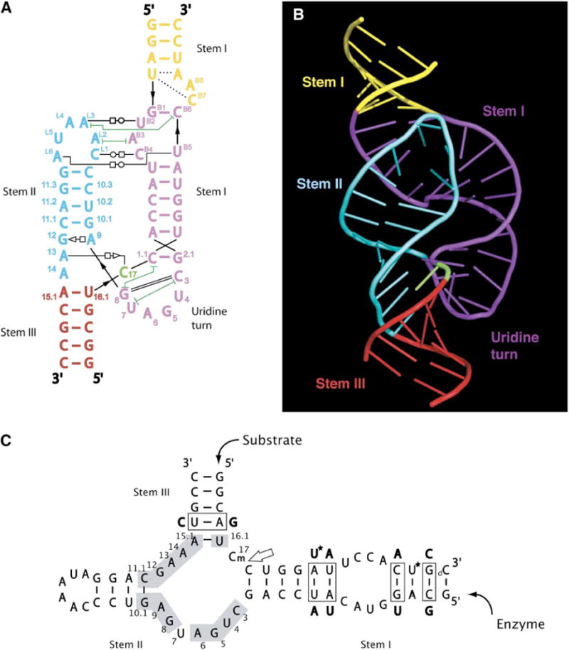
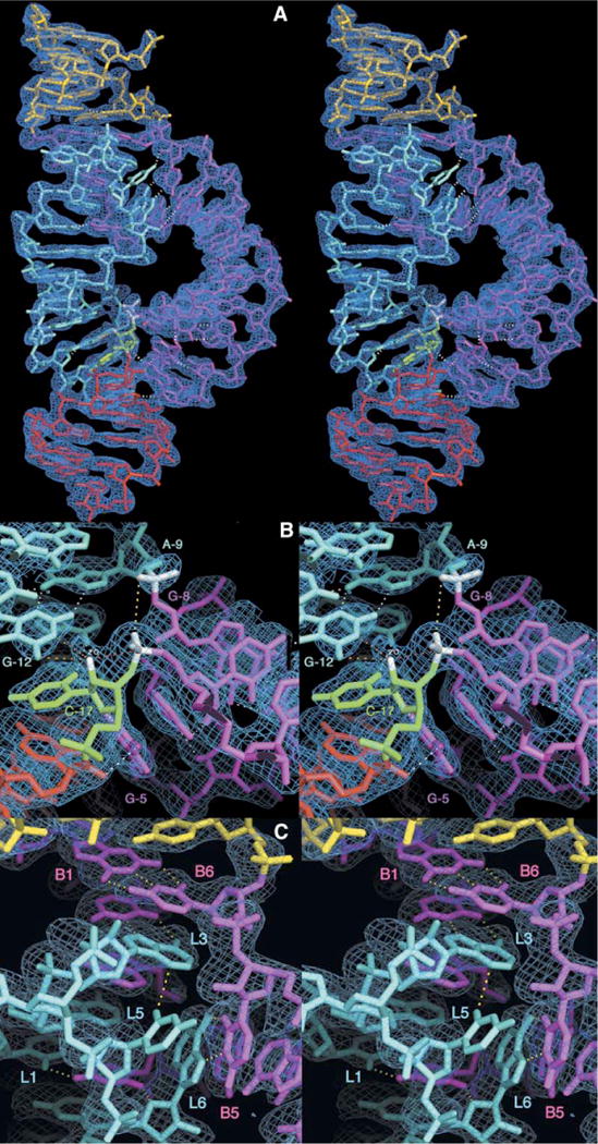
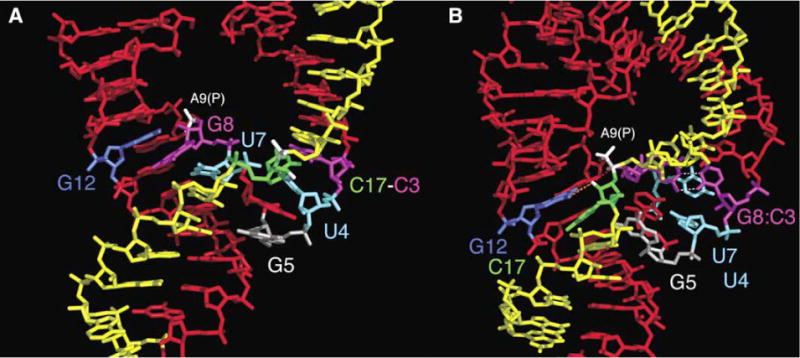
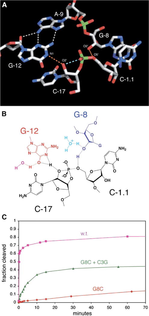
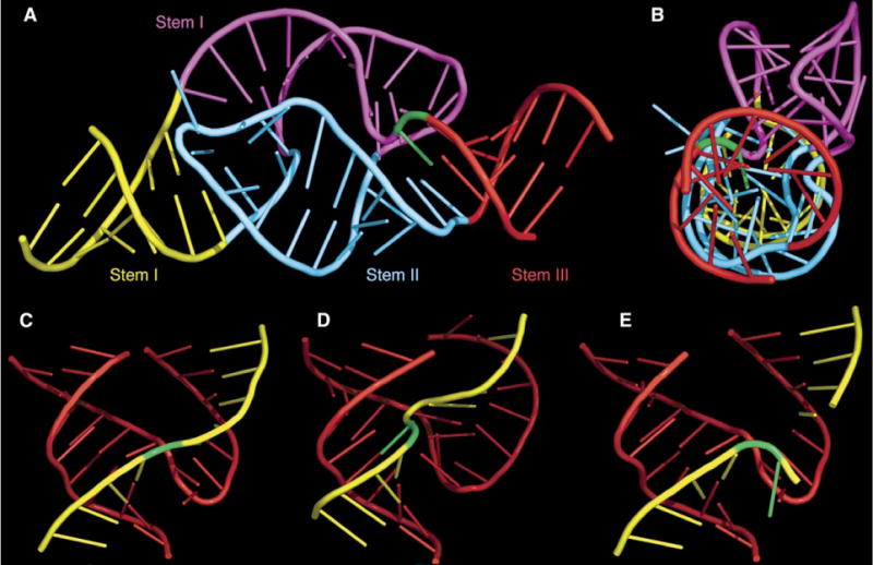
Minimal hammerhead ribozymes have been characterized extensively by static and time-resolved crystallography as well as numerous biochemical analyses, leading to mutually contradictory mechanistic explanations for catalysis. We present the 2.2 A resolution crystal structure of a full-length Schistosoma mansoni hammerhead ribozyme that permits us to explain the structural basis for its 1000-fold catalytic enhancement. The full-length hammerhead structure reveals how tertiary interactions occurring remotely from the active site prime this ribozyme for catalysis. G-12 and G-8 are positioned consistent with their previously suggested roles in acid-base catalysis, the nucleophile is aligned with a scissile phosphate positioned proximal to the A-9 phosphate, and previously unexplained roles of other conserved nucleotides become apparent within the context of a distinctly new fold that nonetheless accommodates the previous structural studies. These interactions permit us to explain the previously irreconcilable sets of experimental results in a unified, consistent, and unambiguous manner.


Figures

Figure 1
Figure 1. The Sequence and the Structure
The secondary structure of the full-length hammerhead ribozyme (A) is oriented and color coded to complement the three-dimensional structural representation (B). Canonical base-pairing interactions are shown as colored lines, and other hydrogen bonding interactions are shown as thin black lines having various annotations according to the following key (Yang et al., 2003): open circle next to open square = Watson-Crick/Hoogsteen; open square next to open triangle = Hoogsteen/sugar edge; dashed line = single hydrogen bond; green lines with T termini = nonadjacent base stacking. Thick black lines indicate backbone continuity where the sequence has been separated for diagrammatic clarity. The sequence used for structural determination, and its derivation from the Schistosoma hammerhead ribozyme, is shown in (C). The conserved nucleotides are numbered using the standard convention and are shown on a gray background. The large arrow points to the scissile bond. Base-pair switches from the wild-type sequence, introduced to aid crystallization, are boxed, and the base substitutions we employed are indicated adjacent to the boxes. Two 5-bromo-uridine substitutions, used for MAD phasing a single crystal, are indicated by asterisks. The lowercase m signifies a 2′-O-methyl modification at C-17 to prevent cleavage. The lowercase d designates a deoxynucleotide used at the 3′ end of the substrate to increase synthetic yields. Figure 1B and all subsequent molecular model figures were made with PyMOL (http://pymol.sourceforge.net/).
Figure 2
Figure 2. Stereo Diagrams of the Full-Length Hammerhead Ribozyme and the Experimentally Phased MAD Electron Density Map
(A) Refined atomic model of the full-length Schistosoma mansoni hammerhead ribozyme superimposed upon the initial 2.2 Å resolution MAD electron density map contoured at 1.25 rmsd. The color scheme follows that introduced in Figure 1. Hydrogen bonds that mediate tertiary interactions between stems I and II and within the active site are shown as dotted white lines. (B) Closeup of the active site with the same electron density map. Several of the nucleotides implicated in catalysis, including G-12, G-8, A-9, and C-17, are labeled. In addition to the white hydrogen bonds, the yellow dotted line indicates the 4.3 Å distance between the A-9 and scissile phosphates, and the red dotted line indicates the direction of in-line attack of the 2′-O of C-17 upon the adjacent scissile phosphate. The nucleophile and the scissile phosphate, as well as the A-9 phosphate, are highlighted in white. The orange dotted lines represent contacts or hydrogen bonds that may be active in catalysis. The extra bit of density surrounding the 2′-O of C-17 corresponds to the 2′-CH3 bound to the 2′-O to prevent the cleavage reaction from taking place. The methyl group has been omitted from the figure to emphasize the additional electron density but is included in the coordinates that have been deposited in the Protein Data Bank (ID code 3ZP8). (C) Closeup of the stem II loop/stem I bulge interaction, showing a hydrogen bonding network summarized in Figure 1A.
Figure 3
Figure 3. Side-by-Side Comparison of the Minimal and Full-Length Hammerhead Ribozyme Structures
Several dramatic changes induced by the loop/bulge tertiary interaction between stems I and II reshape the active site of the full-length hammerhead ribozyme (B) relative to that of the minimal hammerhead (A). The most radical of these are the formation of a base pair between G-8 and C-3 (magenta) and the complete reorientation of the cleavage-site base, C-17 (green). As a result, the adjacent scissile phosphate (highlighted in white) approaches within 4.3 Å of the A-9 phosphate (also in white). The position of G-12 (blue) is similar in both structures, but in the full-length hammerhead, it interacts with the cleavage-site ribose (orange dotted line), whereas G-8 (magenta) rotates from the position occupied in stem II of the minimal structure to pair with C-3 (magenta), creating an extension of stem I. Similarly, U-7 (cyan) in the full-length hammerhead rotates into the space occupied by the base of U-4 (cyan). G-5 (silver) in the uridine turn helps to position C-17 (green) in the full-length structure by forming a hydrogen bond to its furanose oxygen. The differences are illustrated dynamically with an adiabatic morphing between the minimal and full-length active-site structures (see Movie S1 in the Supplemental Data).
Figure 4
Figure 4. Positioning of Nucleotide Functional Groups for Acid-Base Catalysis
(A) Atoms are color coded as follows: carbon, gray; oxygen, red; nitrogen, blue; phosphorus, green. Hydrogen bonds are shown as white and orange dotted lines, and the light blue dotted line shows that the 2′ oxygen, the attacking nucleophile of C-17, is in line with the scissile phosphorus and leaving-group 5′ oxygen. The orange dotted lines indicate hydrogen bonds that are potentially active for acid-base catalysis. (B) A hypothetical transition-state configuration involving G-12 (red) as a general base, G-8 (blue) as a general acid, and the substrate RNA (black). Specific-base catalysis via a water or hydroxide ion (magenta) may also play a role in this mechanism. Similarly, specific-acid catalysis from a water molecule or hydronium ion (cyan) avoids the need to invoke a fully charged alkoxide anion. (C) The G8-C3 base pair is important for catalysis. The fraction of substrate cleaved as a function of time at pH 7.4 for the active sequence and mutations is shown. Pink squares represent the cleavage time course of the crystallographic construct (kobs = 50 min−1, a positive control). Red diamonds represent the activity of the G8C single mutant (kobs < 0.0001, a negative control), which decreases the activity about 500,000-fold. Green triangles represent the activity of the C3G + G8C double mutant (kobs = 0.22 min−1), which rescues the activity of the ribozyme, demonstrating the importance of the base pair.
Figure 5
Figure 5. Aspects of the Full-Length Hammerhead Ribozyme Fold
(A) Stems II and III are almost perfectly coaxial with the distal end of stem I. The remainder of stem I is quite curved and unwound. (B) 90° rotation of the previous view, highlighting the coaxial stacking of all three stems as well as the profoundly distorted and unwound geometry of the remainder of stem I, highlighted in magenta. (C) The region of the fold of the full-length hammerhead ribozyme (A) that corresponds to the previous, truncated hammerhead ribozyme structures. (D and E) Truncated precleavage (D) and postcleavage (E) hammerhead folds are shown for comparison. In each case, the enzyme strand is red; the substrate is yellow; and C-17, the cleavage-site nucleotide, is green.

References

    1. Blount KF, Uhlenbeck OC. The structure-function dilemma of the hammerhead ribozyme. Annu Rev Biophys Biomol Struct. 2005;34:415–440. - PubMed
    1. Brunger AT, Adams PD, Clore GM, DeLano WL, Gros P, Grosse-Kunstleve RW, Jiang JS, Kuszewski J, Nilges M, Pannu NS, et al. Crystallography & NMR system: A new software suite for macromolecular structure determination. Acta Crystallogr D Biol Crystallogr. 1998;54:905–921. - PubMed
    1. Canny MD, Jucker FM, Kellogg E, Khvorova A, Jayasena SD, Pardi A. Fast cleavage kinetics of a natural hammerhead ribozyme. J Am Chem Soc. 2004;126:10848–10849. - PubMed
    1. CCP4 (Collaborative Computational Project Number 4) The CCP4 suite: programs for protein crystallography. Acta Crystallogr D Biol Crystallogr. 1994;50:760–763. - PubMed
    1. Clouet-d’Orval B, Uhlenbeck OC. Hammerhead ribozymes with a faster cleavage rate. Biochemistry. 1997;36:9087–9092. - PubMed

Publication types

Associated data