

Linkage disequilibrium mapping in domestic dog breeds narrows the progressive rod-cone degeneration interval and identifies ancestral disease-transmitting chromosome
- PMID: 16859891
- PMCID: PMC4006154
- DOI: 10.1016/j.ygeno.2006.05.013
Linkage disequilibrium mapping in domestic dog breeds narrows the progressive rod-cone degeneration interval and identifies ancestral disease-transmitting chromosome
Abstract
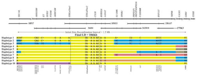
Canine progressive rod-cone degeneration (prcd) is a retinal disease previously mapped to a broad, gene-rich centromeric region of canine chromosome 9. As allelic disorders are present in multiple breeds, we used linkage disequilibrium (LD) to narrow the approximately 6.4-Mb interval candidate region. Multiple dog breeds, each representing genetically isolated populations, were typed for SNPs and other polymorphisms identified from BACs. The candidate region was initially localized to a 1.5-Mb zero recombination interval between growth factor receptor-bound protein 2 (GRB2) and SEC14-like 1 (SEC14L). A fine-scale haplotype of the region was developed, which reduced the LD interval to 106 kb and identified a conserved haplotype of 98 polymorphisms present in all prcd-affected chromosomes from 14 different dog breeds. The findings strongly suggest that a common ancestor transmitted the prcd disease allele to many of the modern dog breeds and demonstrate the power of the LD approach in the canine model.
Conflict of interest statement
Figures
Similar articles
-
Identical mutation in a novel retinal gene causes progressive rod-cone degeneration in dogs and retinitis pigmentosa in humans.Genomics. 2006 Nov;88(5):551-63. doi: 10.1016/j.ygeno.2006.07.007. Epub 2006 Aug 30. Genomics. 2006. PMID: 16938425 Free PMC article.
-
Linkage analysis and comparative mapping of canine progressive rod-cone degeneration (prcd) establishes potential locus homology with retinitis pigmentosa (RP17) in humans.Proc Natl Acad Sci U S A. 1998 Mar 17;95(6):3048-53. doi: 10.1073/pnas.95.6.3048. Proc Natl Acad Sci U S A. 1998. PMID: 9501213 Free PMC article.
-
Assessment of the functionality of genome-wide canine SNP arrays and implications for canine disease association studies.Anim Genet. 2011 Apr;42(2):181-90. doi: 10.1111/j.1365-2052.2010.02132.x. Epub 2010 Nov 11. Anim Genet. 2011. PMID: 21070295
-
Natural models for retinitis pigmentosa: progressive retinal atrophy in dog breeds.Hum Genet. 2019 May;138(5):441-453. doi: 10.1007/s00439-019-01999-6. Epub 2019 Mar 23. Hum Genet. 2019. PMID: 30904946 Review.
-
Dog star rising: the canine genetic system.Nat Rev Genet. 2004 Dec;5(12):900-10. doi: 10.1038/nrg1492. Nat Rev Genet. 2004. PMID: 15573122 Review.
Cited by
-
Exonic SINE insertion in STK38L causes canine early retinal degeneration (erd).Genomics. 2010 Dec;96(6):362-8. doi: 10.1016/j.ygeno.2010.09.003. Epub 2010 Sep 29. Genomics. 2010. PMID: 20887780 Free PMC article.
-
The genetics of canine skull shape variation.Genetics. 2013 Feb;193(2):317-25. doi: 10.1534/genetics.112.145284. Genetics. 2013. PMID: 23396475 Free PMC article.
-
Evaluating the Histologic Grade of Digital Squamous Cell Carcinomas in Dogs and Copy Number Variation of KIT Ligand-A Correlation Study.Vet Sci. 2023 Jan 24;10(2):88. doi: 10.3390/vetsci10020088. Vet Sci. 2023. PMID: 36851392 Free PMC article.
-
Understanding hereditary diseases using the dog and human as companion model systems.Mamm Genome. 2007 Jul;18(6-7):444-51. doi: 10.1007/s00335-007-9037-1. Epub 2007 Jul 26. Mamm Genome. 2007. PMID: 17653794 Free PMC article. Review.
-
Domestic dogs and cancer research: a breed-based genomics approach.ILAR J. 2014;55(1):59-68. doi: 10.1093/ilar/ilu017. ILAR J. 2014. PMID: 24936030 Free PMC article. Review.
References
-
- Peltonen L. Positional cloning of disease genes: advantages of genetic isolates. Hum Hered. 2000;50:66–75. - PubMed
-
- Varilo T, Paunio T, Parker A, Perola M, Meyer J, Terwilliger JD, Peltonen L. The interval of linkage disequilibrium (LD) detected with microsatellite and SNP markers in chromosomes of Finnish populations with different histories. Hum Mol Genet. 2003;12:51–9. - PubMed
Publication types
MeSH terms
Substances
Grants and funding
LinkOut - more resources
Full Text Sources
Other Literature Sources
Research Materials
Miscellaneous
