. 2006 Oct 15;91(8):2848-59.
doi: 10.1529/biophysj.106.084046. Epub 2006 Jul 21.

Interaction of protegrin-1 with lipid bilayers: membrane thinning effect

Affiliations

Interaction of protegrin-1 with lipid bilayers: membrane thinning effect

Hyunbum Jang et al. Biophys J. .

Abstract

Protegrins (PG) are important in defending host tissues, preventing infection via an attack on the membrane surface of invading microorganisms. Protegrins have powerful antibiotic abilities, but the molecular-level mechanisms underlying the interactions of their beta-sheet motifs with the membrane are not known. Protegrin-1 (PG-1) is composed of 18 amino acids with a high content of basic residues and two disulfide bonds. Here we focused on the stability of PG-1 at the amphipathic interface in lipid bilayers and on the details of the peptide-membrane interactions. We simulated all-atom models of the PG-1 monomer with explicit water and lipid bilayers composed of both homogeneous POPC (palmitoyl-oleyl-phosphatidylcholine) lipids and a mixture of POPC/POPG (palmitoyl-oleyl-phosphatidylglycerol) (4:1) lipids. We observed that local thinning of the lipid bilayers mediated by the peptide is enhanced in the lipid bilayer containing POPG, consistent with experimental results of selective membrane targeting. The beta-hairpin motif of PG-1 is conserved in both lipid settings, whereas it is highly bent in aqueous solution. The conformational dynamics of PG-1, especially the highly charged beta-hairpin turn region, are found to be mostly responsible for disturbing the membrane. Even though the eventual membrane disruption requires PG-1 oligomers, our simulations clearly show the first step of the monomeric effects. The thinning effects in the bilayer should relate to pore/channel formation in the lipid bilayer and thus be responsible for further defects in the membrane caused by oligomer.


Figures
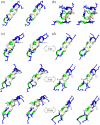
FIGURE 1
FIGURE 1
Cartoons of PG-1 in stereo view, representing (a) the NMR structure, (b) the final peptide conformation from the water box simulation, and snapshots at 1 ns, 5 ns, and 10 ns for the peptides from (c) the pure bilayer system and (d) the mixed bilayer system. In the cartoons, hydrophobic residues and disulfide-bonded Cys residues are shown in white, a polar residue (Tyr) and Gly are shown in green, and positively charged residues (Arg) are shown in blue. Disulfide bonds are highlighted in yellow.
FIGURE 2
FIGURE 2
The average RMSD from the starting point for Cα atoms (upper panel) and Cβ atoms (lower panel) of PG-1 in the water box (open squares), on the pure lipid bilayer composed of POPC (inverted gray triangles), and on the mixed lipid bilayer composed of POPC/POPG (4:1) (solid circles).
FIGURE 3
FIGURE 3
(a) Probability of interaction energies with the surrounding environments for PG-1 in the water box (upper panel), on the pure lipid bilayer composed of POPC (middle panel), and on the mixed lipid bilayer composed of POPC/POPG (4:1) (lower panel). (b) The interaction energy with the lipid is separated into POPC (upper panel) and POPG (lower panel) lipid parts for the mixed lipid bilayer system. The total interaction energies as well as the electrostatic contributions and the vdW contributions are shown.
FIGURE 4
FIGURE 4
Time series of peptide displacement from the average position of phosphate atoms on the extracellular side for PG-1 on the pure lipid bilayer composed of POPC (black line) and the mixed lipid bilayer composed of POPC/POPG (4:1) (gray line).
FIGURE 5
FIGURE 5
(a) Probability distribution functions (P) for different component groups of lipid (PChol (choline), PPO4 (phosphate), PGlyc (glycerol), PCarb (carbonyls), and PCH3 (methyl)) and for other solvents (Pwater (water), PNa (sodium ion), and PCl (chloride ion)) as a function of distance from the bilayer center for PG-1 on the pure lipid bilayer composed of POPC. (b) The same probability distribution functions for PG-1 on the mixed lipid bilayer composed of POPC/POPG (4:1). The probability distribution functions are separated into POPC (upper panel) and POPG (middle panel) lipid parts. In the distribution function for POPG, PChol (choline) from POPC is replaced byformula image (top-head glycerol). (c) Highlight of the probability distribution function for water, Pwater, for the pure lipid bilayer (red), and for the mixed lipid bilayer systems (blue). PG-1 is located at the lipid/water interface in the positive z region.
FIGURE 6
FIGURE 6
Deuterium order parameters, SCD, for the oleoyl (left column) and palmitoyl (right) chains for the different lipid categories, for the extracellular (peptide-containing) side (upper panel) and the intracellular (lower) side, and for the pure lipid bilayer composed of POPC (all circles) and the mixed lipid bilayer composed of POPC/POPG (4:1) (all triangles). The local lipids (those adjacent to the peptide) are denoted by open symbols (open circles and triangles), and the bulk lipids are denoted by filled symbols (solid circles and gray triangles).
FIGURE 7
FIGURE 7
Mapping of x, y coordinates of the center of mass of each lipid on the extracellular half lipids onto the x-y plane for (a) the pure lipid bilayer composed of POPC and (b) the mixed lipid bilayer composed of POPC/POPG (4:1). The contour lines enclose highly populated locations of the center of mass of each POPC (gray lines) and POPG (black lines). The peptide is also projected onto the plane as an average structure with its average position during the simulation. Hydrophobic residues including Gly and disulfide-bonded Cys residues are shown in white, a polar residue (Tyr) is shown in green, and positively charged residues (Arg) are shown in blue.
FIGURE 8
FIGURE 8
Three-dimensional density maps of the lipid headgroup for (a) the pure lipid bilayer composed of POPC and (b) the mixed lipid bilayer composed of POPC/POPG (4:1). In both figures, the upper figure is the top view from the extracellular side with an embedded peptide and the lower figure is the lateral view of the bilayer. In the lateral bilayer views, the blue lines are the three-point connection that measures the bilayer bending, and the vertical double arrow points the degree of the bilayer thickness. Note that the three points and lines are only guides to eyes.
FIGURE 9
FIGURE 9
Three-dimensional density map of water (blue), sodium (yellow), and chloride (white) ions for the pure lipid bilayer composed of POPC (upper) and the mixed lipid bilayer composed of POPC/POPG (4:1) (lower). In the peptide, hydrophobic residues including Gly and disulfide-bonded Cys residues are shown in white, a polar residue (Tyr) is shown in green, and positively charged residues (Arg) are shown in blue.

References

    1. Kourie, J. I., and A. A. Shorthouse. 2000. Properties of cytotoxic peptide-formed ion channels. Am. J. Physiol. Cell Physiol. 278:C1063–C1087. - PubMed
    1. Lai, J. R., B. R. Huck, B. Weisblum, and S. H. Gellman. 2002. Design of non-cysteine-containing antimicrobial β-hairpins: structure-activity relationship studies with linear protegrin-1 analogues. Biochemistry. 41:12835–12842. - PubMed
    1. Gobbo, M., L. Biondi, F. Filira, R. Gennaro, M. Benincasa, B. Scolaro, and R. Rocchi. 2002. Antimicrobial peptides: synthesis and antibacterial activity of linear and cyclic drosocin and apidaecin 1b analogues. J. Med. Chem. 45:4494–4504. - PubMed
    1. Panchal, R. G., M. L. Smart, D. N. Bowser, D. A. Williams, and S. Petrou. 2002. Pore-forming proteins and their application in biotechnology. Curr. Pharm. Biotechnol. 3:99–115. - PubMed
    1. Sitaram, N., and R. Nagaraj. 1999. Interaction of antimicrobial peptides with biological and model membranes: structural and charge requirements for activity. Biochim. Biophys. Acta. 1462:29–54. - PubMed

Publication types

MeSH terms

LinkOut - more resources