. 2006 Aug 1;103(31):11515-20.
doi: 10.1073/pnas.0603921103. Epub 2006 Jul 21.

Regulation of neddylation and deneddylation of cullin1 in SCFSkp2 ubiquitin ligase by F-box protein and substrate

Affiliations

Regulation of neddylation and deneddylation of cullin1 in SCFSkp2 ubiquitin ligase by F-box protein and substrate

Gil Bornstein et al. Proc Natl Acad Sci U S A. .

Abstract

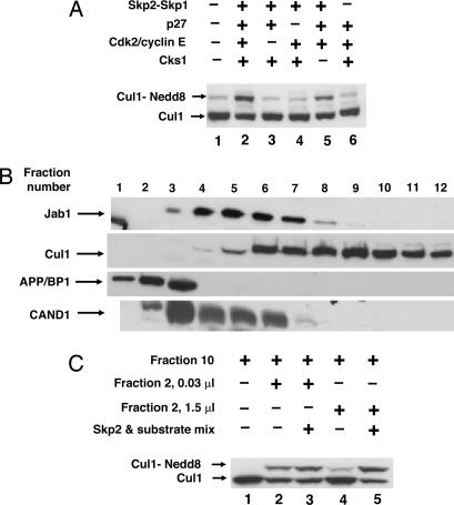
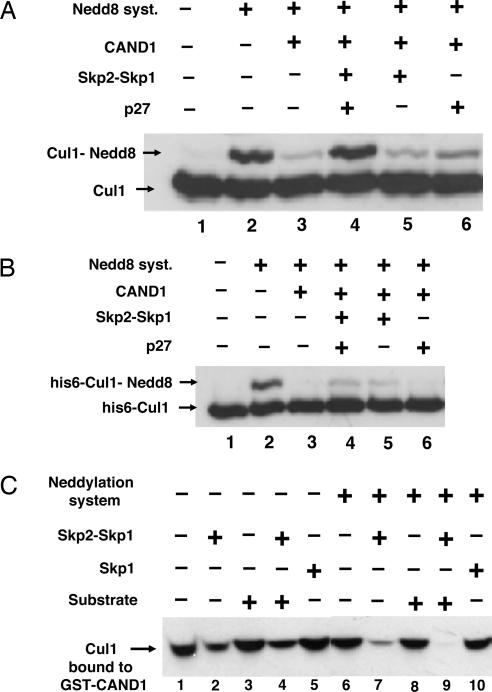
The activity of cullin-containing ubiquitin protein ligase complexes is stimulated by linkage to cullin of the ubiquitin-like protein Nedd8 ("neddylation"). Neddylation is inhibited by the tight binding of cullins to CAND1 (cullin-associated and neddylation-dissociated 1) protein, and Nedd8 is removed from cullins by specific isopeptidase activity of the COP9/signalosome (CSN) complex. The mechanisms that regulate neddylation and deneddylation of cullins were unknown. We examined this problem for the case of SCF(Skp2), a cullin1 (Cul1)-containing ubiquitin ligase complex that contains the S phase-associated protein Skp2 as the substrate-binding F-box protein subunit. SCF(Skp2) targets for degradation the cyclin-dependent kinase (cdk) inhibitor p27 in the G(1)-to-S phase transition, a process that requires its phosphorylation and binding to cdk2-cyclin E. Because levels of Skp2, cyclin E, and the accessory protein Cks1 (cyclin kinase subunit 1) all rise at the end of G(1) phase, it seemed possible that the neddylation of Cul1 in SCF(Skp2) is regulated by the availability of the F-box protein and/or the substrate. We found that the supplementation of Skp2-Skp1 and substrate (along with further components necessary for substrate presentation to the ubiquitin ligase) to extracts of HeLa cells synergistically increased levels of neddylated Cul1. Skp2-Skp1 abrogates the inhibitory influence of CAND1 on the neddylation of Cul1 by promoting the dissociation of the cullin-CAND1 complex, whereas substrate, together with substrate-presenting components, prevents the action of CSN to deneddylate cullin. We propose a sequence of events in which the increased availability of Skp2 and substrate in the transition of cells to S phase promotes the neddylation and assembly of the SCF(Skp2) ubiquitin ligase complex.


Conflict of interest statement

Conflict of interest statement: No conflicts declared.

Figures

Fig. 1.
Fig. 1.
Skp2 and substrate synergistically increase levels of neddylated Cul1 in extracts and fractions from HeLa cells. (A) Increase in levels of neddylated Cul1 in extracts of HeLa cells by combined addition of Skp2–Skp1, p27 substrate, and other components required for the interaction of substrate with SCFSkp2. The neddylation of endogenous Cul1 in extracts of HeLa cells (3 μg of protein) was assayed as described in Materials and Methods, except that the concentration of APP-BP1-Uba3 was 0.01 nM. Skp2–Skp1 was added as indicated in the presence of either complete substrate mixture (lane 2) or substrate mixtures from which the indicated components were omitted (lanes 3–5). (B) Fractionation of HeLa cell extracts on a heparin-Sepharose column. Extracts of HeLa cells were fractionated as described in Materials and Methods. Samples of 3 μl of the indicated column fractions were subjected to immunoblotting with the indicated antibodies. (C) Reconstitution of the effects of Skp2 and substrate with fractions of extract separated on a heparin-Sepharose column. The effects of Skp2 and substrate on the neddylation of Cul1 were assayed as described in Materials and Methods, except that recombinant APP-BP1-Uba3 was not added. Samples (0.5 μl) of fraction 10 of the heparin column served as the source of Cul1. The indicated amounts of fraction 2 from the same heparin column were added as specified.
Fig. 2.
Fig. 2.
Skp2 causes dissociation of Cul1 from CAND1 and, jointly with substrate, abrogates inhibition of neddylation. (A) Skp2 and substrate overcome inhibition by CAND1 of the neddylation of Cul1 in a partially purified preparation. Assay of the neddylation of Cul1 was carried out as described in Materials and Methods in the presence of 0.5 μl of fraction 9 of the heparin column as the source of Cul1. In lanes 3 and 5, a substrate mixture lacking p27 was added. Where indicated, CAND1 (70 nM) was added 5 min before the neddylation mixture. (B) Neddylation of purified recombinant Cul1 in the presence of CAND1 is only slightly stimulated by Skp2–Skp1. Experimental conditions were as in A, except that partially purified Cul1 was replaced by 35 nM recombinant purified His-6-Cul1-Roc1. (C) Skp2–Skp1, but not Skp1 or substrate, promotes dissociation of Cul1–Cand1 complex. Bacterially expressed GST–CAND1 (1.3 pmol) was preincubated with 4 μl of pooled fractions 10–12 from the heparin-Sepharose column for 30 min at 30°C in a buffer consisting of 50 mM Tris·HCl, pH 7.6/5 mM MgCl2/1 mM DTT/10% glycerol/10 mM phosphocreatine/0.1 μg/μl creatine phosphokinase/0.5 mM ATP/3 mg/ml BSA. Subsequently, Skp2–Skp1 (10 nM), Skp1 (15 nM), or complete substrate mixture (see Materials and Methods) was added as indicated, and a second incubation (at 20°C for 15 min) was carried out under conditions as described for the assay of neddylation of Cul1, except that the reaction volume was 40 μl and a neddylation system (consisting of APP-BP1-Uba3, Ubc12, and Nedd8) was added only in the indicated tubes. Next, 10 μl of glutathione-Sepharose beads (Amersham Pharmacia Biosciences) was added, and samples were rotated at 4°C for 90 min. The beads were washed three times with a buffer consisting of 50 mM Tris·HCl, pH 7.2/1 mM DTT/2 mg/ml BSA/100 mM NaCl/10% glycerol, eluted with SDS sample buffer, and subjected to immunoblotting for Cul1 for the estimation of Cul1 that remained bound to GST–CAND1.
Fig. 3.
Fig. 3.
Substrate prevents the deneddylation of Cul1 by CSN. (A) The neddylation of affinity-purified Cul1 bound to GST–CAND1 is stimulated by Skp2 in the absence of substrate, and the requirement for substrate is restored by the fraction not adsorbed to GST–CAND1. Samples of 0.5 μl of affinity-purified Cul1 from HeLa cells bound to GST–CAND1 (see Materials and Methods) were subjected to the neddylation assay. Where indicated, 0.5 μl of the fraction not adsorbed to GST–CAND1, Skp2, or substrate mixture containing or lacking p27 was supplemented. (B) Experimental conditions were as in A except that the unadsorbed fraction was replaced by the indicated amounts of purified CSN (see Materials and Methods). (C) Substrate prevents the deneddylation of Cul1 (in complex with Skp2–Skp1) by CSN. The first incubation contained in a volume of 7 μl: 50 mM Tris·HCl (pH 7.6), 10% glycerol, 20 nM Skp2–Skp1, 0.5 μl of affinity-purified Cul1 in complex with GST–CAND1, and neddylation mixture as described above. After incubation at 20°C for 30 min, a mixture of 0.1 μg/μl hexokinase, 20 mM 2-deoxyglucose, and 20 mM DTT was added to stop the neddylation reaction. After 5 min, 0.05 μl of purified CSN was added in the presence (lane 4) or absence (lane 3) of complete substrate mixture. Substrate was added 5 min before CSN. After a second incubation (at 20°C for 30 min), samples were subjected to electrophoresis and immunoblotting with antibody to Cul1.
Fig. 4.
Fig. 4.
Proposed sequence of events in the neddylation and assembly of the SCFSkp2 complex. See the text (Discussion). The increase in the levels of Skp2–Skp1 in the transition of cells to S phase promotes the dissociation of Cul1 from CAND1 and thus allows its neddylation. The increase in the levels of substrate in the same stage of the cell cycle locks the SCFSkp2 complex in the neddylated and assembled state by preventing deneddylation. “Substrate” denotes phosphorylated p27 in ternary complex with cdk2-cyclin E and bound to Cks1.

References

    1. Petroski M. D., Deshaies R. J. Nat. Rev. Mol. Cell Biol. 2005;6:9–20. - PubMed
    1. Cardozo T., Pagano M. Nat. Rev. Mol. Cell Biol. 2004;5:739–751. - PubMed
    1. Carrano A. C., Eytan E., Hershko A., Pagano M. Nat. Cell Biol. 1999;1:193–199. - PubMed
    1. Ganoth D., Bornstein G., Ko T. K., Larsen B., Tyers M., Pagano M., Hershko A. Nat. Cell Biol. 2001;3:321–324. - PubMed
    1. Kawakami T., Chiba T., Suzuki T., Iwai K., Yamanaka K., Minato N., Suzuki H., Shimbara N., Hidaka Y., Osaka F., et al. EMBO J. 2001;20:4003–4012. - PMC - PubMed

Publication types

MeSH terms