. 2006 Aug;74(8):4452-61.
doi: 10.1128/IAI.00666-06.

Construction and characterization of an attenuated purine auxotroph in a Francisella tularensis live vaccine strain

Affiliations

Construction and characterization of an attenuated purine auxotroph in a Francisella tularensis live vaccine strain

Roger Pechous et al. Infect Immun. 2006 Aug.

Abstract

Francisella tularensis is a facultative intracellular pathogen and is the etiological agent of tularemia. It is capable of escaping from the phagosome, replicating to high numbers in the cytosol, and inducing apoptosis in macrophages of a variety of hosts. F. tularensis has received significant attention recently due to its potential use as a bioweapon. Currently, there is no licensed vaccine against F. tularensis, although a partially protective live vaccine strain (LVS) that is attenuated in humans but remains fully virulent for mice was previously developed. An F. tularensis LVS mutant deleted in the purMCD purine biosynthetic locus was constructed and partially characterized by using an allelic exchange strategy. The F. tularensis LVS delta purMCD mutant was auxotrophic for purines when grown in defined medium and exhibited significant attenuation in virulence when assayed in murine macrophages in vitro or in BALB/c mice. Growth and virulence defects were complemented by the addition of the purine precursor hypoxanthine or by introduction of purMCDN in trans. The F. tularensis LVS delta purMCD mutant escaped from the phagosome but failed to replicate in the cytosol or induce apoptotic and cytopathic responses in infected cells. Importantly, mice vaccinated with a low dose of the F. tularensis LVS delta purMCD mutant were fully protected against subsequent lethal challenge with the LVS parental strain. Collectively, these results suggest that F. tularensis mutants deleted in the purMCD biosynthetic locus exhibit characteristics that may warrant further investigation of their use as potential live vaccine candidates.


Figures

FIG. 1.
FIG. 1.
PCR and Southern blot analyses of four constructed F. tularensis LVS ΔpurMCD mutants. (A) Genomic organization of the purMCD region in F. tularensis LVS. NdeI restriction enzyme sites are present within the coding sequences for folD and purCD and in the beginning of the groE-aph kanamycin resistance gene. Primers used for PCR confirmation of mutants lie outside the cloned region and are indicated with arrows. The thick solid line indicates DNA from the purMCD upstream region used as the probe for Southern blotting. (B) PCR products resulting from amplification of F. tularensis LVS or F. tularensis LVS ΔpurMCD::groE-aph genomic DNA with primers purCDFlankF1 and purCDFlankR1. DNA (2.2 kb) between folD and purN was removed following deletion of purMCD and replacement with groE-aph. (C) Southern blot of total genomic DNA from wild-type LVS or ΔpurMCD::groE-aph mutants digested with NdeI and hybridized to radiolabeled probe from the purMCD upstream region. Replacement of purMCD with groE-aph reduces the size of the hybridized band from 3.6 kb to 1.0 kb due to the presence of an NdeI site within groE-aph. L, ladder; M1 to M4, mutants 1 to 4.
FIG.2.
FIG.2.
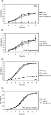
Growth kinetics of F. tularensis LVS derivatives in defined and rich media. Wild-type F. tularensis LVS, the ΔpurMCD mutant, and the complemented ΔpurMCD mutant were grown in CDM in the absence (A) or presence (B) of 50 μg/ml purine precursor hypoxanthine or in modified MH broth medium lacking (C) or containing (D) 2.5% fetal bovine serum and 1.0% proteose peptone. Growth was monitored by measuring the optical density at 550 nm (OD 550nm). The means and standard errors of experiments performed in triplicate are shown.
FIG. 3.
FIG. 3.
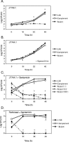
Intracellular growth of F. tularensis LVS derivatives in macrophages. J774A.1 macrophage-like cells (A to C) or peritoneal macrophages (D) were infected with wild-type F. tularensis LVS, the ΔpurMCD mutant, and the complemented ΔpurMCD mutant, and intracellular growth was monitored by lysing macrophages at the indicated time points and determining CFU. All infections were conducted at an MOI of 1 unless noted otherwise. Gentamicin (5 μg/ml) was added to culture medium after infection (C and D) to kill extracellular organisms that were not ingested or that were released from infected macrophages. Asterisks indicate time points where growth of the F. tularensis LVS ΔpurMCD mutant was significantly different (P < 0.05; ANOVA) from growth of LVS or the genetic complement. The means and standard errors of experiments performed in triplicate are shown.
FIG. 4.
FIG. 4.
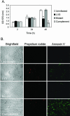
Effect of F. tularensis LVS growth on macrophage adherence. (A) Retention of crystal violet stain was used as an indicator of adherence of J774A.1 macrophages infected with F. tularensis LVS derivatives. A significant drop (asterisks, P < 0.05; ANOVA) in adherence was observed 48 h after infection in macrophages infected (MOI of 10) with wild-type LVS or the complemented mutant. No differences in staining were observed between the F. tularensis ΔpurMCD mutant and the uninfected controls. OD 570 (nm), optical density at 570 nm. (B) Apoptosis in F. tularensis-infected J774A.1 macrophages was examined using a Vibrant apoptosis assay kit no. 2 to stain annexin V and propidium iodide to measure permeability. Macrophages were seeded onto coverslips in tissue culture dishes and were left uninfected or infected with wild-type F. tularensis LVS, the ΔpurMCD mutant, or the complemented mutant for 48 h. Infected cells were washed, stained with annexin V conjugated to Alexa Fluor 488 and red fluorescent propidium iodide, and mounted onto glass slides for analysis by epifluorescence microscopy. Note the absence of annexin V staining in macrophages infected with the F. tularensis ΔpurMCD mutant. Representative images from experiments performed in triplicate are shown.
FIG. 5.
FIG. 5.
Intracellular localization of F. tularensis LVS derivatives inside macrophages. Murine BMMs were infected with F. tularensis LVS derivatives and analyzed by TEM (A) or epifluorescence microscopy following Francisella and LAMP-1 immunostaining (B). BMMs were infected with F. tularensis derivatives at an MOI of 50 (for TEM) or 25 (for epifluorescence microscopy) and processed at 2 h and 8 h postinfection (pi). The F. tularensis LVS ΔpurMCD mutant escapes from the phagosome but is unable to replicate in the cytosol of infected macrophages. The F. tularensis LVS ΔpurMCD mutant bacteria are also found at low frequency in LAMP-1-positive phagosomes in infected macrophages. LAMP-1 colocalization was determined for each strain by counting 100 bacteria at each time point. Values are expressed as the means ± SD from experiments preformed in triplicate. Bar, 0.5 μm.
FIG. 6.
FIG. 6.
Growth characteristics of F. tularensis LVS derivatives in BALB/c mice. Groups of mice were infected intraperitoneally with wild-type LVS (50 CFU), the ΔpurMCD mutant (48 CFU), or the complemented mutant (26 CFU). At specific times after infection, subsets of mice were sacrificed and the numbers of bacteria present in the spleens (A) or livers (B) were determined by plating organ homogenates on MH agar medium. Values depicted at each time point represent the mean log CFU per ml ± standard errors of the means from groups of three animals. The F. tularensis LVS ΔpurMCD mutant was not recovered from spleen or liver tissues at any time examined. The remaining subset of infected animals was used to define the mean time required by each bacterial strain to induce morbidity (C). Percent survival was determined from groups of 10 mice per infecting bacterial strain.

References

    1. Brown, R. F., and B. A. D. Stocker. 1987. Salmonella typhi 205aTy, a strain with two attenuating auxotrophic characters, for use in laboratory teaching. Infect. Immun. 55:892-898. - PMC - PubMed
    1. Burke, D. S. 1977. Immunization against tularemia: analysis of the effectiveness of live Francisella tularensis vaccine in prevention of laboratory-acquired tularemia. J. Infect. Dis. 135:55-60. - PubMed
    1. Chamberlain, R. E. 1965. Evaluation of live tularemia vaccine prepared in a chemically defined medium. Appl. Microbiol. 13:232-235. - PMC - PubMed
    1. Crawford, R. M., L. Van De Verg, L. Yuan, T. L. Hadfield, R. L. Warren, E. S. Drazek, H. H. Houng, C. Hammack, K. Sasala, T. Polsinelli, J. Thompson, and D. L. Hoover. 1996. Deletion of purE attenuates Brucella melitensis infection in mice. Infect. Immun. 64:2188-2192. - PMC - PubMed
    1. Eigelsbach, H. T., and C. M. Downs. 1961. Prophylactic effectiveness of live and killed tularemia vaccines. I. Production of vaccine and evaluation in the white mouse and guinea pig. J. Immunol. 87:415-425. - PubMed

Publication types

MeSH terms

LinkOut - more resources