

The presence of peptidoglycan O-acetyltransferase in various staphylococcal species correlates with lysozyme resistance and pathogenicity
- PMID: 16861647
- PMCID: PMC1539615
- DOI: 10.1128/IAI.00301-06
The presence of peptidoglycan O-acetyltransferase in various staphylococcal species correlates with lysozyme resistance and pathogenicity
Abstract
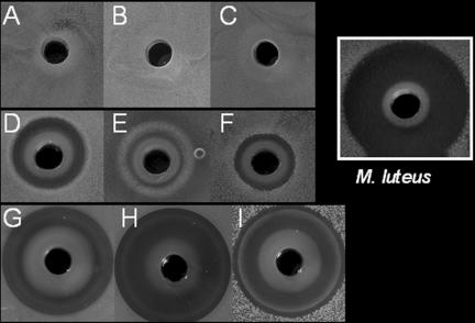
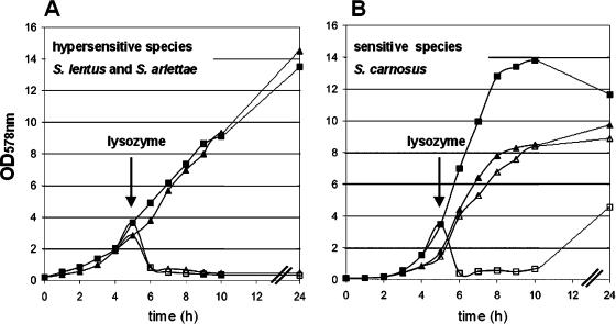
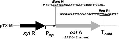
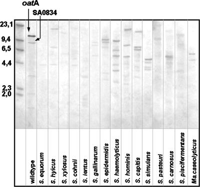
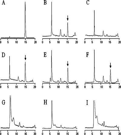
Human-pathogenic bacteria that are able to cause persistent infections must have developed mechanisms to resist the immune defense system. Lysozyme, a cell wall-lytic enzyme, is one of the first defense compounds induced in serum and tissues after the onset of infection. Recently, we showed that Staphylococcus aureus is resistant to lysozyme by O acetylating its peptidoglycan (PG) by O-acetyltransferase (OatA). We asked the question of which staphylococcal species PG is O acetylated. We applied various methods, such as genome analysis, PCR, Southern blotting, lysozyme sensitivity assay, and verification of O acetylation of PG by high-performance liquid chromatography (HPLC) analysis. PCR analysis using S. aureus-derived oatA primers and Southern blotting did not yield reliable results with other staphylococcal species. Therefore, we used the HPLC-based assay to directly detect PG O acetylation. Our studies revealed that the muramic acid was O acetylated only in pathogenic, lysozyme-resistant staphylococci (e.g., S. aureus, S. epidermidis, S. lugdunensis, and others). All nonpathogenic species were lysozyme sensitive. They can be divided into sensitive species (e.g., S. carnosus, S. gallinarum, and S. xylosus) and hypersensitive species (e.g., S. equorum, S. lentus, and S. arlettae). In all lysozyme-sensitive species, the analyzed PG was de-O-acetylated. When we transformed the oatA gene from lysozyme-resistant S. aureus into S. carnosus, the corresponding transformants also became lysozyme resistant.
Figures
References
-
- Archer, G. L. 1998. Staphylococcus aureus: a well-armed pathogen. Clin. Infect. Dis. 26:1179-1181. - PubMed
-
- Baneyx, F., and M. Mujacic. 2004. Recombinant protein folding and misfolding in Escherichia coli. Nat. Biotechnol. 22:1399-1408. - PubMed
-
- Bartoszewicz, M., J. Nowicka, and A. Przondo-Mordarska. 2003. Selected features determine pathogenicity of Staphylococcus haemolyticus. Med. Dosw. Mikrobiol. 55:225-229. - PubMed
-
- Bera, A., S. Herbert, A. Jakob, W. Vollmer, and F. Gotz. 2005. Why are pathogenic staphylococci so lysozyme resistant? The peptidoglycan O-acetyltransferase OatA is the major determinant for lysozyme resistance of Staphylococcus aureus. Mol. Microbiol. 55:778-787. - PubMed
-
- Blaiotta, G., C. Pennacchia, F. Villani, A. Ricciardi, R. Tofalo, and E. Parente. 2004. Diversity and dynamics of communities of coagulase-negative staphylococci in traditional fermented sausages. J. Appl. Microbiol. 97:271-284. - PubMed
Publication types
MeSH terms
Substances
LinkOut - more resources
Full Text Sources
Medical
Molecular Biology Databases
