. 2007 Feb 1;26(5):757-64.
doi: 10.1038/sj.onc.1209828. Epub 2006 Jul 24.

ATR-dependent checkpoint modulates XPA nuclear import in response to UV irradiation

Affiliations

ATR-dependent checkpoint modulates XPA nuclear import in response to UV irradiation

X Wu et al. Oncogene. .

Abstract

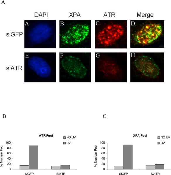
In response to DNA damage, mammalian cells activate various DNA repair pathways to remove DNA lesions and, meanwhile, halt cell cycle progressions to allow sufficient time for repair. The nucleotide excision repair (NER) and the ATR-dependent cell cycle checkpoint activation are two major cellular responses to DNA damage induced by UV irradiation. However, how these two processes are coordinated in the response is poorly understood. Here we showed that the essential NER factor XPA (xeroderma pigmentosum group A) underwent nuclear accumulation upon UV irradiation, and strikingly, such an event occurred in an ATR (Ataxia-Telangiectasia mutated and RAD3-related)-dependent manner. Either treatment of cells with ATR kinase inhibitors or transfection of cells with small interfering RNA targeting ATR compromised the UV-induced XPA nuclear translocation. Consistently, the ATR-deficient cells displayed no substantial XPA nuclear translocation while the translocation remained intact in ATM (Ataxia-Telangiectasia mutated)-deficient cells in response to UV irradiation. Moreover, we found that ATR is required for the UV-induced nuclear focus formation of XPA. Taken together, our results suggested that the ATR checkpoint pathway may modulate NER activity through the regulation of XPA redistribution in human cells upon UV irradiation.


Figures

Figure 1
Figure 1. Induction of XPA nuclear accumulation by UV irradiation
(A) Exponentially growing A549 cells were treated with increasing doses of UV followed by a 2 h recovery. Proteins from the nuclear extracts, N, and cytoplasmic fractions, C, of the cells were separated on SDS-PAGE gels and probed with anti-XPA antibody (left panel). The UV-treated whole cells were also analyzed by immunofluorescence microscopy (middle panel). The nuclei were stained with DAPI (4',6-diamidino-2-phenylindole). Specificity of anti-XPA antibody used in these experiments and the total levels of XPA in the whole cell lysates versus UV doses were demonstrated by Western blotting in the right panel. GADPH was used as the loading control. (B) A549 cells were treated with 10 J/m2 UV irradiation, and then harvested at the indicated time after treatment. Cytoplasmic and nuclear extracts were prepared and probed with anti-XPA antibody. The UV-treated whole cells were also analyzed by immunofluorescence microscopy in a time-dependent manner (right panel). (C) A549 cells were treated with indicated doses of camptothecin (CPT) for 2 h, and then washed with PBS and further incubated for another 2 h. Cytoplasmic and nuclear extracts were prepared and probed with anti-XPA antibody.
Figure 2
Figure 2. UV-induced XPA nuclear import is wortmannin and caffeine sensitive
(A) A549 cells were mocked treated or treated with 20 J/m2 UV irradiation, and then incubated for 4 h in the presence of 100 μM wortmannin (Wort) (lanes 5 and 6) or 10 mM caffeine (Caff) (lanes 3 and 4) before harvesting. Nuclear extracts were analyzed by Western blotting probed with anti-XPA, anti-ATR and anti-ATM, respectively. Asterisk represents the phosphorylated form of XPA, which is also wortmannin and caffeine sensitive (Wu et al., 2006). (B) The UV-induced XPA nuclear accumulation was quantified using densitometry. The values (the mean ± SD of three independent experiments) were normalized to that for mock treated cells (as the value of 1).
Figure 3
Figure 3. ATR is required for XPA nuclear accumulation following UV irradiation
(A) A549 cells were transfected with indicated siRNA, and then treated with 20 J/m2 UV 72 h after transfection. Nuclear extracts were isolated 4 h after UV treatment and immunoblotted with the antibodies indicated above. Asterisk represents the phosphorylated form of XPA (Wu et al., 2006). (B)The quantities of nuclear XPA after UV treatment were estimated using densitometry and normalized to those obtained from mock treated cells, which were designated as 1.0. (C) A549 cells were transfected with siRNA targeting either ATR or GFP, or mock-transfected and then irradiated with 20 J/m2 UV 72 hours after transfection. Cells were then fixed and stained with antibodies for either XPA (red) or calreticulin (green), an ER-lumen protein; nuclei were stained with DAPI. Subpanels c and f illustrate the cellular distribution of XPA in normal (mock-transfected) cells with XPA located primarily in the cytosol prior to UV irradiation (subpanel c) and accumulating in the nuclei following irradiation (subpanel f). Cells transfected with siRNA for ATR did not show similar translocation of XPA to the nucleus following UV irradiation (subpanel i). However, cells transfected with GFP siRNA had no effect on XPA nuclear accumulation (subpanel l). (D) Removal of UV-induced (6-4)PPs over time were assayed in the cells transfected with siRNAs for ATR and GFP, respectively, as analyzed by immunofluorescence with anti-(6-4)PPs antibody. The UV was irradiated through a filter containing 5 μm pores overlaid on the cells. The foci represent the (6-4)PPs lesions.
Fig. 4
Fig. 4. UV-induced XPA nuclear accumulation is defective in ATR deficient cells
(A) A549 cells, ATR and ATM deficient cells were treated with indicated doses of UV, and nuclear extracts were prepared 4 h after treatment for Western blotting with anti-XPA antibody. Asterisk represents the phosphorylated form of XPA (Wu et al., 2006). (B) The quantitative data of Figure 4A, which represent the mean ± SD of three independent experiments.
Fig. 5
Fig. 5. ATR is required for the UV-induced XPA foci formation
(A) A549 cells were transfected with ATR siRNA or GFP siRNA as described in Materials and Methods, and then treated with 20 J/m2 UV, 72 h after transfection. Cells were then subjected to immunofluorescence assays with anti-XPA and anti-ATR antibodies. Subpanels B and F (green) are anti-XPA stained cells; Subpanels C and G (red) are anti-ATR stained cells. Subpanels D and H are the merged images of the anti-XPA and anti-ATR stained cells. Subpanels A and E are DAPI (4',6-diamidino-2-phenylindole)-stained nuclei. (B) and (C) The UV-induced ATR and XPA nuclear foci were scored from a total of randomly picked at least 100 cells in two independent experiments. The cells containing more than 10 foci/cell were defined as positively stained cells. The data represented the percentage of positively stained cells.

References

    1. Abraham RT. Cell cycle checkpoint signaling through the ATM and ATR kinases. Genes Dev. 2001;15:2177–96. - PubMed
    1. Adimoolam S, Ford JM. p53 and DNA damage-inducible expression of the xeroderma pigmentosum group C gene. Proc Natl Acad Sci U S A. 2002;99:12985–90. - PMC - PubMed
    1. Barr SM, Leung CG, Chang EE, Cimprich KA. ATR kinase activity regulates the intranuclear translocation of ATR and RPA following ionizing radiation. Curr Biol. 2003;13:1047–51. - PubMed
    1. Bartek J, Lukas C, Lukas J. Checking on DNA damage in S phase. Nat Rev Mol Cell Biol. 2004;5:792–804. - PubMed
    1. Cliby WA, Roberts CJ, Cimprich KA, Stringer CM, Lamb JR, Schreiber SL, et al. Overexpression of a kinase-inactive ATR protein causes sensitivity to DNA-damaging agents and defects in cell cycle checkpoints. Embo J. 1998;17:159–69. - PMC - PubMed

Publication types

MeSH terms

Substances