

The effect of lithium on the adrenoceptor-mediated second messenger system in the rat brain
- PMID: 16862242
- PMCID: PMC1488903
The effect of lithium on the adrenoceptor-mediated second messenger system in the rat brain
Abstract
Objective: Lithium remains the most widely used treatment for bipolar disorder; however, the molecular mechanisms underlying its therapeutic actions have not been fully elucidated. We studied the in-vivo effect of lithium on the density of alpha-adrenoceptor (alpha-AR) and beta-AR subtypes and linked second messenger systems in the rat brain.
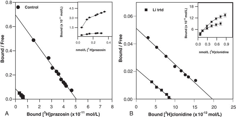
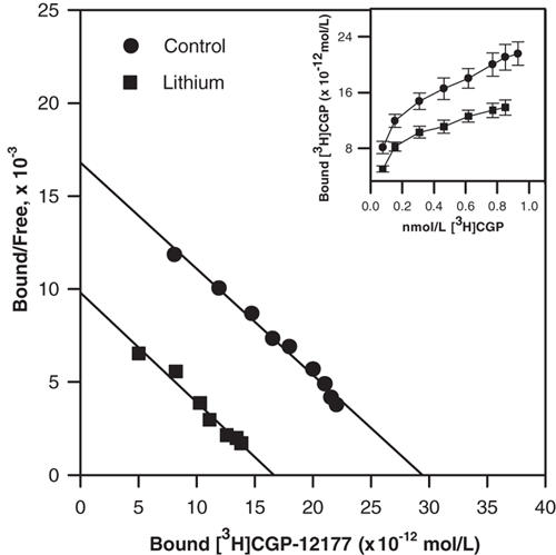
Methods: The densities of alpha(1)-ARs, alpha(2)-ARs, and beta(1)-ARs and beta(2)-ARs in the cortex and cerebellum of rats treated with lithium (0.4%), orally, for 30 days were measured using [(3)H]prazosin, [(3)H]clonidine and [(3)H]CGP-12177, respectively. The activity of adenylyl cyclase (AC) and levels of inositol trisphosphate (IP3), both second messengers linked to these receptors, were estimated using [(3)H]ATP and [(3)H]myoinositol, respectively.
Results: A significant decrease in the densities of cortical alpha(1)-ARs (85%, p < 0.0001), alpha(2)-ARs (50%, p < 0.0001), beta(1)-ARs (26%, p < 0.0001) and beta(2)-ARs (25%, p < 0.0001) was observed after lithium treatment. However, only the density of alpha(1)-ARs was significantly decreased (25%, p < 0.0001) in the cerebellum. The affinity of [(3)H]prazosin for cerebellar alpha(1)-ARs was increased. A small, but statistically significant, increase (19%, p < 0.0001) in the density of total beta-ARs was seen in the cerebellum, without altering the affinity of the radioligand for these receptors. Basal AC activity was not altered in the lithium-treated rat cortex. However, the norepinephrine-stimulated AC activity, which represents alpha(2)-AR-linked and beta-AR-linked AC, was significantly increased (66%, p < 0.0001). Both basal IP3 formation and norepinephrine-stimulated IP3, which represents alpha(1)-AR-linked phospholipase C activity, were significantly decreased (50%, p < 0.0001) in the lithium-treated rat cortex.
Conclusion: Our results suggest that long-term administration of lithium treatment downregulates the cortical, but not cerebellar, alpha(1)-ARs, alpha(2)-ARs, beta(1)-ARs and beta(2)-ARs. Thus, it may be concluded that lithium induces region-specific and differential functional downregulation of alpha-AR and beta-AR subtypes in the rat brain.
Objectif: Le lithium demeure le traitement le plus répandu du trouble bipolaire, mais on n'a pas élucidé entièrement les mécanismes moléculaires qui en sous-tendent les effets thérapeutiques. Nous avons étudié l'effet in vivo du lithium sur la densité des sous-types α-adrénorécepteur (α-AR) et β-AR et des systèmes de second messager reliés dans le cerveau du rat.
Méthodes: On a mesuré les densités des récepteurs α1-AR, α2-AR, β1-AR et β2-AR dans le cortex et le cervelet de rats traités au lithium (0,4 %) par voie orale pendant 30 jours en utilisant la [3H]prazosine, la [3H]clonidine et le [3H]CGP-12177, respectivement. On a estimé l'activité de l'adénylyl cyclase (AC) et les concentrations d'inositol trisphosphate (IP3), tous deux seconds messagers reliés à ces récepteurs, en utilisant [3H]ATP et [3H]myoinositol respectivement.
Résultats: On a observé, après le traitement au lithium, une diminution importante des densités d'α1-AR (85 %, p < 0,0001), d'α2-AR (50 %, p < 0,0001), de β1-AR (26 %, p < 0,0001) et de β2-AR (25 %, p < 0,0001) dans le cortex. Dans le cervelet, toutefois, seule la densité de α1-AR a diminué considérablement (25 %, p < 0,0001). L'affinité de la [3H]prazosine pour les α1-AR du cervelet a augmenté. On a constaté une augmentation modeste mais statistiquement significative (19 %, p < 0,0001) de la densité du total des β-AR dans le cervelet sans qu'il y ait modification de l'affinité des radioligands pour ces récepteurs. L'activité basale de l'AC n'a pas changé dans le cortex des rats traités au lithium. L'activité de l'AC stimulée par la norépinéphrine, qui représente l'AC reliée aux récepteurs α2-AR et au β-AR, a toutefois augmenté considérablement (66 %, p < 0,0001). La formation d'IP3 basal et d'IP3 stimulé par la norépinéphrine, qui représente l'activité de la phospholipase C reliée aux α1-AR, a toutefois diminué considérablement (50 %, p < 0,0001) dans le cortex des rats traités au lithium.
Conclusion: Nos résultats indiquent que l'administration d'un traitement de longue durée au lithium entraîne une régulation à la baisse des α1-AR, α2-AR, β1-AR et β2-AR dans le cortex, mais non dans le cervelet. On peut donc conclure que le lithium produit une régulation à la baisse fonctionnelle, différentielle et spécifique à une région des sous-types α-AR et β-AR dans le cerveau du rat.
Figures
Similar articles
-
Differential modulation of α-1 adrenoceptor subtypes by antidepressants in the rat brain.J Neural Transm (Vienna). 2010 Dec;117(12):1423-30. doi: 10.1007/s00702-010-0522-4. Epub 2010 Dec 7. J Neural Transm (Vienna). 2010. PMID: 21136124
-
Effect of amitriptyline on adrenergic receptor number and second messenger function in rat brain.Pak J Biol Sci. 2012 Sep 15;15(18):871-6. doi: 10.3923/pjbs.2012.871.876. Pak J Biol Sci. 2012. PMID: 24205756
-
Differential spatiotemporal alterations in adrenoceptor mRNAs and binding sites in cerebral cortex following spreading depression: selective and prolonged up-regulation of alpha1B-adrenoceptors.Exp Neurol. 1998 Dec;154(2):612-27. doi: 10.1006/exnr.1998.6915. Exp Neurol. 1998. PMID: 9878196
-
Are so many adrenergic receptor subtypes really present in domestic animal tissues? A pharmacological perspective.Vet J. 2005 Sep;170(2):163-74. doi: 10.1016/j.tvjl.2004.05.015. Epub 2004 Oct 22. Vet J. 2005. PMID: 16129337 Review.
-
Signal transduction pathways. Molecular targets for lithium's actions.Arch Gen Psychiatry. 1995 Jul;52(7):531-43. doi: 10.1001/archpsyc.1995.03950190013003. Arch Gen Psychiatry. 1995. PMID: 7598629 Review.
Cited by
-
What is the Role of Lithium in Epilepsy?Curr Neuropharmacol. 2022;20(10):1850-1864. doi: 10.2174/1570159X20666220411081728. Curr Neuropharmacol. 2022. PMID: 35410603 Free PMC article. Review.
-
IP3 accumulation and/or inositol depletion: two downstream lithium's effects that may mediate its behavioral and cellular changes.Transl Psychiatry. 2016 Dec 6;6(12):e968. doi: 10.1038/tp.2016.217. Transl Psychiatry. 2016. PMID: 27922641 Free PMC article.
-
Differential modulation of α-1 adrenoceptor subtypes by antidepressants in the rat brain.J Neural Transm (Vienna). 2010 Dec;117(12):1423-30. doi: 10.1007/s00702-010-0522-4. Epub 2010 Dec 7. J Neural Transm (Vienna). 2010. PMID: 21136124
References
-
- Ahlquist RP. A study of the adrenotropic receptors. Am J Physiol 1948;153:586-600. - PubMed
-
- Langer SZ. Presynaptic regulation of catecholamine release. Biochem Pharmacol 1974;23:1793-800. - PubMed
-
- Lands AM, Arnold A, McAuliff JP, et al. Differentiation of receptor systems activated by sympathomimetic amines. Nature 1967;214: 597-8. - PubMed
-
- Berridge MJ, Irvin RF. Inositol phosphates and cell signaling. Nature 1989;341:197-205. - PubMed
-
- Hauger RL, Rehavi M, Angel I, et al. Receptor-mediated mechanism of antidepressants drug action in psychiatry. In: Michef R, Cavanar JD, editors. Psychobiological foundation of clinical psychiatry. Vol. 3. Philadelphia: Lippincott; 1998. p. 1-22.
Publication types
MeSH terms
Substances
LinkOut - more resources
Full Text Sources
Research Materials