

doi: 10.1016/j.pnpbp.2006.06.007.
Epub 2006 Jul 24.
Volumetric alterations of the orbitofrontal cortex in autism
Affiliations
- PMID: 16863674
- PMCID: PMC2888006
- DOI: 10.1016/j.pnpbp.2006.06.007
Item in Clipboard
Volumetric alterations of the orbitofrontal cortex in autism
Prog Neuropsychopharmacol Biol Psychiatry.
.
Abstract
Recent evidence has implicated the orbitofrontal cortex (OFC) in the pathophysiology of social deficits in autism. An MRI-based morphometric study of the OFC was conducted involving 11 children with autism (age range 8.1-12.7 years) and 18 healthy, age-matched controls (age range 8.9-12.8 years). Decreased grey matter volume in the right lateral OFC in the patient group was found, and correlations were observed between social deficits and white, but not grey, matter structures of the OFC. These findings support the role of OFC in autism and warrant further investigations of this structure using structural and functional methodologies.
Figures
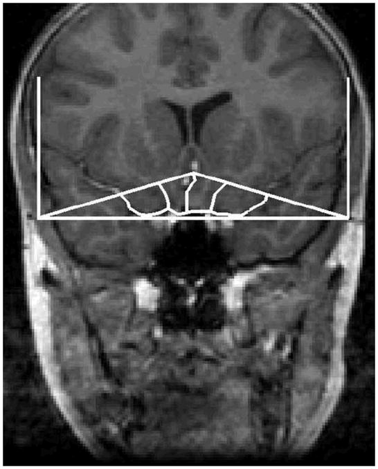
A tracing of the orbitofrontal cortex in the coronal plane.
References
-
- Abell F, Krams M, Ashburner J, Passingham R, Friston K, Frackowiak R, Happe F, Frith C, Frith U. The neuroanatomy of autism: a voxel-based whole brain analysis of structural scans. Neuroreport. 1999;10:1647–1651. - PubMed
-
- Aylward EH, Minshew NJ, Field K, Sparks BF, Singh N. Effects of age on brain volume and head circumference in autism. Neurology. 2002;59:175–183. - PubMed
-
- Bachevalier J, Loveland KA. The orbitofrontal-amygdala circuit and self regulation of social-emotional behavior in autism. Neurosci Biobehav Rev. 2006;30:97–117. - PubMed
-
- Barnea-Goraly N, Kwon H, Menon V, Eliez S, Lotspeich L, Reiss AL. White matter structure in autism: preliminary evidence from diffusion tensor imaging. Biol Psychiatry. 2004;55:323–326. - PubMed
-
- Boddaert N, Chabane N, Gervais H, Good CD, Bourgeois M, Plumet MH, Barthelemy C, Mouren MC, Artiges E, Samson Y, Brunelle F, Frackowiak RS, Zilbovicius M. Superior temporal sulcus anatomical abnormalities in childhood autism: a voxel-based morphometry MRI study. Neuroimage. 2004;23:364–369. - PubMed
Publication types
MeSH terms
Grants and funding
LinkOut - more resources
Full Text Sources
