. 2006 Jul 31;174(3):425-36.
doi: 10.1083/jcb.200604031. Epub 2006 Jul 24.

Transport of PIP3 by GAKIN, a kinesin-3 family protein, regulates neuronal cell polarity

Affiliations

Transport of PIP3 by GAKIN, a kinesin-3 family protein, regulates neuronal cell polarity

Kaori Horiguchi et al. J Cell Biol. .

Abstract

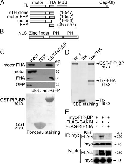
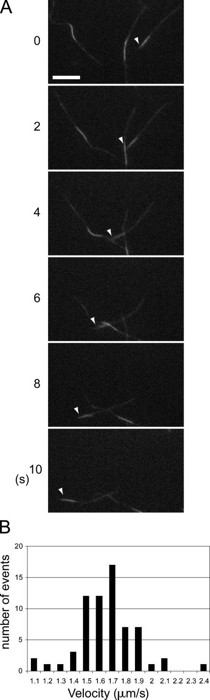
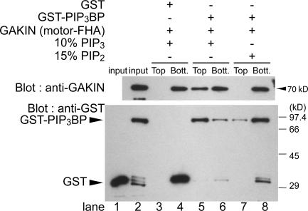
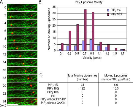
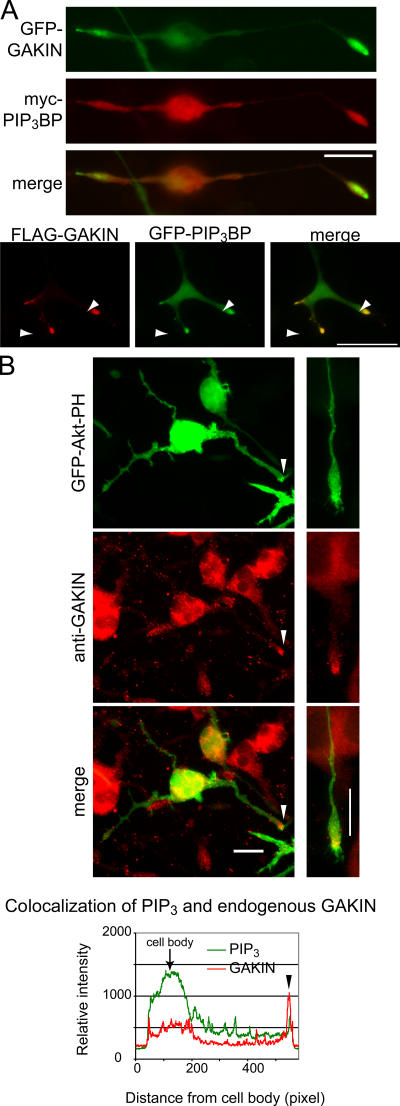
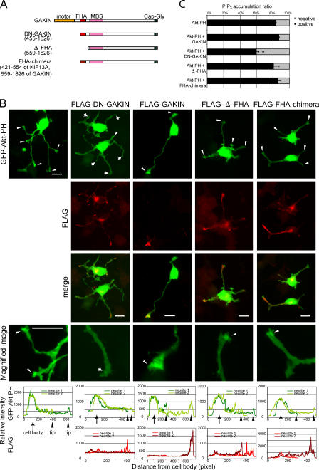
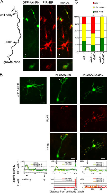
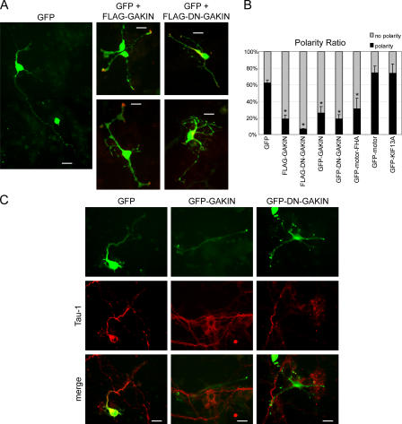
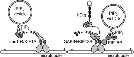
Phosphatidylinositol-(3,4,5)-trisphosphate (PIP3), a product of phosphatidylinositol 3-kinase, is an important second messenger implicated in signal transduction and membrane transport. In hippocampal neurons, the accumulation of PIP3 at the tip of neurite initiates the axon specification and neuronal polarity formation. We show that guanylate kinase-associated kinesin (GAKIN), a kinesin-like motor protein, directly interacts with a PIP3-interacting protein, PIP3BP, and mediates the transport of PIP3-containing vesicles. Recombinant GAKIN and PIP3BP form a complex on synthetic liposomes containing PIP3 and support the motility of the liposomes along microtubules in vitro. In PC12 cells and cultured hippocampal neurons, transport activity of GAKIN contributes to the accumulation of PIP3 at the tip of neurites. In hippocampal neurons, altered accumulation of PIP3 by overexpression of GAKIN constructs led to the loss of the axonally differentiated neurites. Together, these results suggest that, in neurons, the GAKIN-PIP3BP complex transports PIP3 to the neurite ends and regulates neuronal polarity formation.


Figures

Figure 1.
Figure 1.
The FHA domain of GAKIN mediates the biochemical interaction with PIP3BP. (A) Schematic representation of GAKIN domain organization and various constructs. Kinesin family motor domain (motor), FHA, MAGUK binding stalk domain (MBS), and microtubule binding CAP-Gly domain are depicted. YTH indicates the clone obtained from the yeast two-hybrid screen. (B) Domain organization of PIP3BP. Nuclear localization signal (NLS), Arf-GAP–like zinc-finger motif (zinc finger), and PH domains are depicted. (C) GST pull-down assay. Defined segments of GAKIN were expressed as GFP fusion proteins in 293T cells, and their interactions with PIP3BP were tested by GST pull-down assay using GST-PIP3BP. GFP-GAKIN segments pulled down by GST-PIP3BP were detected by Western blotting using anti-GFP polyclonal antibody. Recombinant GST and GST-PIP3BP proteins used for the pull-down experiments were shown by Ponceau staining of the nitrocellulose membrane. (D) Direct interaction between purified GST-PIP3BP and Trx-FHA of GAKIN. Trx-FHA and control Trx proteins were immobilized on S-protein agarose beads (Novagen) and incubated with GST-PIP3BP. Beads were recovered by centrifugation, and bound proteins were analyzed by SDS-PAGE and Coomassie blue staining. (E) Coimmunoprecipitation of PIP3BP and GAKIN. COS-7 cells were transfected with plasmids encoding full-length clones of FLAG-GAKIN or -KIF13A and myc-PIP3BP. Immunoprecipitation was performed from the cell lysates using anti-myc monoclonal antibody. FLAG- and myc-tagged proteins were detected by Western blotting using anti-FLAG and anti-myc monoclonal antibodies.
Figure 2.
Figure 2.
GAKIN is a microtubule plus-end motor. (A) Microtubules gliding on the glass surface coated with motor-FHA of GAKIN. Microtubules were polarity labeled by Alexa 488 to highlight their minus ends. Arrowheads point to the minus end of the microtubule. Motility was observed on glass coverslips coated with GAKIN motor-FHA (1–557) recombinant protein. Microtubules moved toward their minus ends with a velocity of ∼1.66 μm/s, suggesting that GAKIN is a plus-end motor. Bar, 5 μm. (B) Histogram of the velocity of microtubules gliding.
Figure 3.
Figure 3.
PIP3BP and GAKIN form a complex on synthetic liposomes containing PIP3. Binding of recombinant GST-PIP3BP and GAKIN motor-FHA (1–557) to liposomes was examined using a membrane flotation assay. PIP3-containing liposomes (10% PIP3 and 90% PC) or PIP2-containing liposomes (15% PIP2 and 85% PC) were incubated with GAKIN motor-FHA (1–557) and GST-PIP3BP or control GST, and liposome and soluble protein fractions were separated by sucrose gradient centrifugation. Liposomes were recovered from the top of sucrose step gradient, whereas the unbound protein remained at the bottom. GAKIN motor-FHA (1–557) and GST-PIP3BP in the top and bottom fractions were detected by Western blotting using anti-GAKIN and anti-GST polyclonal antibodies, respectively.
Figure 4.
Figure 4.
GAKIN transports PIP3 liposomes via PIP3BP in vitro. (A) Time-lapse visualization of a rhodamine-labeled PIP3 liposome (red) moving along microtubules (green) in the presence of GST-PIP3BP and GAKIN motor-FHA (1–557) recombinant proteins. Bar, 5 μm. (B) Histogram of the velocity of liposome motility generated by GAKIN motor-FHA (1–557) at 18 mM ATP (see Materials and methods). (C) Specificity of liposome motility by recombinant GAKIN motor-FHA (1–557) with or without PIP3 and GST-PIP3BP. Liposome motility was observed only in the presence of PIP3, recombinant GST-PIP3BP, GAKIN, and ATP.
Figure 5.
Figure 5.
PIP3, PIP3BP, and GAKIN colocalize at the tip of growing neurites in PC12 cells. (A) PIP3BP and GAKIN colocalize at the tip of growing neurites and growth cones of PC12 cells. PC12 cells were transfected with GFP-PIP3BP (full length) and FLAG-GAKIN (full length) or GFP-GAKIN and myc-PIP3BP. 1 d after transfection, cells were stimulated with 5.0 ng/ml NGF for 36 h and fixed for immunofluorescence analysis. The merged image shows significant colocalization at the distal end of the neurite. Higher magnification images of cells transfected with GFP-PIP3BP and FLAG-GAKIN show significant colocalization of PIP3BP and GAKIN at the tip of the growth cone. (B) PIP3 colocalizes with endogenous GAKIN at the tip of growing neurites. PC12 cells were transfected with GFP-Akt-PH to monitor PIP3 localization and stimulated with NGF as described. Endogenous GAKIN was stained with anti-GAKIN monoclonal antibody. The merged image and measured relative fluorescence intensity show significant colocalization at the tip of neurites.
Figure 6.
Figure 6.
GAKIN contributes to the accumulation of PIP3 at the tip of growing neurites in PC12 cells. (A) Schematic diagram of domain organization of several GAKIN constructs used in these experiments. (B) PIP3 accumulation is blocked by DN-GAKIN. PC12 cells were transfected with GFP-Akt-PH and FLAG-DN-GAKIN, FLAG-GAKIN (full length), FLAG-Δ-FHA, or FLAG-FHA-chimera and stimulated with NGF. Arrowheads represent neurites with PIP3 accumulation at the tip, whereas arrows represent neurites without PIP3 accumulation. Overexpression of DN-GAKIN significantly reduced the accumulation of PIP3 at the neurite tips. Bars, 20 μm. (C) Quantification of PIP3 accumulation at the neurite tips. The numbers of the neurites with and without accumulation of PIP3 were counted. Values from three independent experiments are represented as means + SD (percentage of PIP3 accumulation: GFP-Akt-PH alone, 84 ± 0.5%, n = 1007; GAKIN + GFP-Akt-PH, 87 ± 0.7%, n = 395, P = 0.002; DN-GAKIN + GFP-Akt-PH, 60 ± 3%, n = 693, P < 0002; Δ-FHA + GFP-Akt-PH, 81 ± 7%, n = 220, P = 0.5; FHA-chimera + GFP-Akt-PH, 86 ± 4%, n = 131, P = 0.4; *, P < 0.002).
Figure 7.
Figure 7.
PIP3 and PIP3BP colocalize at the tip of the axon in hippocampal neurons, and this accumulation is modulated by the overexpression of GAKIN. (A) PIP3 colocalizes with endogenous PIP3BP at the tip of the axon in cultured mouse hippocampal neurons. Hippocampal neurons were transfected with GFP-Akt-PH. Endogenous PIP3BP was stained with anti-PIP3BP rabbit antiserum. Arrowheads point to the tip of the axon. Magnified images are shown in the bottom panels. (left) Diagram shows the outline of the cell observed. (B) Overexpression of GAKIN constructs alters the PIP3 localization. Hippocampal neurons were transfected with GFP-Akt-PH and FLAG-GAKIN or FLAG-DN-GAKIN. Overexpression of full-length GAKIN enhanced the accumulation of PIP3, often at the multiple termini of the neurites. Overexpression of DN-GAKIN caused the loss of accumulation of PIP3 at the end of the axon-like neurite. (C) The subpopulation of hippocampal neurons scored as relative fluorescence intensity of GFP-Akt-PH as (neurite tip/cell body) ratio. Fluorescence intensity at the tip was measured and normalized by the intensity at the cell body. The population of cells that show high accumulation at the tip (tip/cell body ratio >1), normal accumulation (tip/cell body ratio >0.5 to <1) or less accumulation (tip/cell body ratio <0.5) are shown in the graphical format (number of cell analyzed and value of a t test: GFP-Akt-PH, n = 23; DN-GAKIN + GFP-Akt-PH, n = 31, P < 0.01; GAKIN + GFP-Akt-PH, n = 27, P < 0.01).
Figure 8.
Figure 8.
GAKIN-mediated transport of PIP3 regulates neuronal polarity of hippocampal neurons. (A) Overexpression of GAKIN constructs affects the neuronal polarity. GFP, as a morphological marker, was cotransfected with FLAG-GAKIN or FLAG-DN-GAKIN. Neuronal polarity was determined as described in Materials and methods. Overexpression of GAKIN, as well as DN-GAKIN, caused the loss of the axonally specified neurites. (B) Quantification of neuronal polarity formation as determined in A (percentage of neurons polarized: GFP, 62 ± 3%, n = 185; GFP-GAKIN, 26 ± 8%, n = 131, P < 0.002; GFP-DN-GAKIN, 19 ± 5%, n = 193, P < 0.0005; GFP-motor, 74 ± 8%, n = 55, P = 0.08; GFP-motor-FHA, 31 ± 13%, n = 43, P = 0.02; GFP-KIF13A, 74 ± 11%, n = 32, P = 0.16; FLAG-GAKIN, 19 ± 4%, n = 120, P < 0.0002: FLAG-DN-GAKIN, 6.4 ± 1%, n = 185, P < 0.00002; *, P < 0.05). (C) Loss of the Tau-1–positive axon structure by overexpression of GAKIN and DN-GAKIN. Bars, 20 μm.
Figure 9.
Figure 9.
A proposed model for the intracellular transport mechanism of PIP2 and PIP3 cargo vesicles by kinesins.

Similar articles

Cited by

References

    1. Aoki, K., T. Nakamura, K. Fujikawa, and M. Matsuda. 2005. Local phosphatidylinositol 3,4,5-trisphosphate accumulation recruits Vav2 and Vav3 to activate Rac1/Cdc42 and initiate neurite outgrowth in nerve growth factor-stimulated PC12 cells. Mol. Biol. Cell. 16:2207–2217. - PMC - PubMed
    1. Asaba, N., T. Hanada, A. Takeuchi, and A.H. Chishti. 2003. Direct interaction with a kinesin-related motor mediates transport of mammalian discs large tumor suppressor homologue in epithelial cells. J. Biol. Chem. 278:8395–8400. - PubMed
    1. Banker, G., and K. Goslin. 1998. Culturing Nerve Cells. 2nd edition. The MIT Press, Cambridge, MA. 666 pp.
    1. Bourne, H.R., and O. Weiner. 2002. A chemical compass. Nature. 419:21. - PubMed
    1. Cantley, L.C. 2002. The phosphoinositide 3-kinase pathway. Science. 296:1655–1657. - PubMed

Publication types

MeSH terms