. 2006 Aug 1;103(31):11784-9.
doi: 10.1073/pnas.0604681103. Epub 2006 Jul 24.

Glatiramer acetate fights against Alzheimer's disease by inducing dendritic-like microglia expressing insulin-like growth factor 1

Affiliations

Glatiramer acetate fights against Alzheimer's disease by inducing dendritic-like microglia expressing insulin-like growth factor 1

Oleg Butovsky et al. Proc Natl Acad Sci U S A. .

Abstract

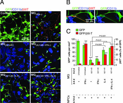
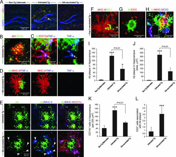
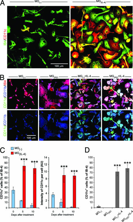
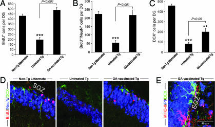
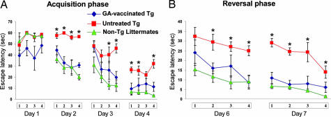
Alzheimer's disease (AD) is characterized by plaque formation, neuronal loss, and cognitive decline. The functions of the local and systemic immune response in this disease are still controversial. Using AD double-transgenic (APP/PS1) mice, we show that a T cell-based vaccination with glatiramer acetate, given according to a specific regimen, resulted in decreased plaque formation and induction of neurogenesis. It also reduced cognitive decline, assessed by performance in a Morris water maze. The vaccination apparently exerted its effect by causing a phenotype switch in brain microglia to dendritic-like (CD11c) cells producing insulin-like growth factor 1. In vitro findings showed that microglia activated by aggregated beta-amyloid, and characterized as CD11b(+)/CD11c(-)/MHC class II(-)/TNF-alpha(+) cells, impeded neurogenesis from adult neural stem/progenitor cells, whereas CD11b(+)/CD11c(+)/MHC class II(+)/TNF-alpha(-) microglia, a phenotype induced by IL-4, counteracted the adverse beta-amyloid-induced effect. These results suggest that dendritic-like microglia, by facilitating the necessary adjustment, might contribute significantly to the brain's resistance to AD and argue against the use of antiinflammatory drugs.


Conflict of interest statement

Conflict of interest statement: No conflicts declared.

Figures

Fig. 1.
Fig. 1.
IL-4 can counteract the adverse effect of aggregated Aβ on microglial toxicity and induces neurogenesis in adult mouse neural progenitor cells. (A) Representative confocal microscopic images of NPCs expressing GFP and βIII-T cocultured for 10 days without microglia (control), or with untreated microglia, or with microglia that were preactivated by aggregated Aβ(1–40) (5 μM) [MG(Aβ1–40)] for 48 h and subsequently activated with IFN-γ (10 ng/ml) [MG(Aβ1–40/IFNγ,10 ng/ml)] or with IL-4 (10 ng/ml) [MG(Aβ1–40/IL-4)] or with both IFN-γ (10 ng/ml) and IL-4 (10 ng/ml) [MG(Aβ1–40/IFNγ+IL-4)]. Note that aggregated Aβ induced microglia to adopt an amoeboid morphology, but after IL-4 was added they exhibited a ramified structure. (B) Separate confocal images of NPCs coexpressing GFP and βIII-T adjacent to CD11b+ microglia. (C) Quantification of cells double-labeled with GFP and βIII-T (expressed as a percentage of GFP+ cells) obtained from confocal images. Results are of three independent experiments in replicate cultures; bars represent means ± SEM. Asterisks above bars denote the significance of differences relative to untreated (control) NPCs (∗, P < 0.05; ∗∗∗, P < 0.001; two-tailed Student’s t test). Horizontal lines with P values above them show differences between the indicated groups (ANOVA).
Fig. 2.
Fig. 2.
GA vaccination induces expression of CD11c by microglia and leads to a reduction in accumulation of Aβ in the brains of Tg-AD mice. (A) Representative confocal microscopic images of brain hippocampal slices from non-Tg, untreated Tg-AD, and GA-vaccinated Tg-AD mice stained for CD11b (activated microglia), human Aβ counterstained with nuclear DAPI. The non-Tg mouse shows no staining for human Aβ. The untreated Tg-AD mouse shows an abundance of extracellular Aβ plaques, whereas in the GA-treated Tg-AD mouse Aβ immunoreactivity is low. There is a high incidence of microglia double-stained for Aβ and CD11b in the CA1 and dentate gyrus regions of the hippocampus of an untreated Tg-AD mouse, but only a minor presence of CD11b+ microglia in the GA-vaccinated Tg-AD mouse. (B) High magnification of CD11b+ microglia associated with an Aβ plaque in an untreated Tg-AD mouse (arrow in A). (C) CD11b+ microglia, associated with an Aβ plaque, strongly expressing TNF-α in an untreated Tg-AD mouse. (D) Staining for MHC-II in GA-vaccinated Tg-AD mouse in an area that stained positively for Aβ shows a high incidence of MHC-II+ microglia and almost no TNF-α+ microglia. (E) All MHC-II+ microglia in a brain area that stained positively for Aβ (arrowheads) in a GA-vaccinated Tg-AD mouse coexpress CD11c, but only a few CD11c+/MHC-II+ microglia are seen in a corresponding area in the brain of an untreated Tg-AD mouse. (F) MHC-II+ microglia in a GA-vaccinated Tg-AD mouse coexpress IGF1. (G and H) CD3+ T cells are seen in close proximity to an Aβ plaque (G) and are associated with MHC-II+ microglia (H). The boxed area in H shows high magnification of an immunological synapse between a T cell (CD3+) and a microglial cell expressing MHC-II. (I) Histogram showing the total number of Aβ plaques (in a 30-μm hippocampal slice). (J) Histogram showing staining for Aβ immunoreactivity. Note the significant differences between GA-vaccinated Tg-AD and untreated Tg-AD mice, verifying the decreased presence of Aβ plaques in the vaccinated mice. (K) Histogram showing a marked reduction in cells stained for CD11b in the GA-vaccinated Tg-AD mice relative to untreated Tg-AD mice. Note the increase in CD11b+ microglia with age in the non-Tg littermates. (L) Histogram showing significantly more CD3+ cells associated with an Aβ plaque in GA-vaccinated Tg-AD mice than in untreated Tg-AD mice. Quantification of CD3+ cells was analyzed from 30–50 plaques of each mouse tested in this study. Error bars indicate means ± SEM. ∗, P < 0.05; ∗∗∗, P < 0.001 versus non-Tg littermates (Student’s t test). The P value represents a comparison by ANOVA. All of the mice in all groups were included in the analysis (six to eight sections per mouse).
Fig. 3.
Fig. 3.
IL-4 induces microglia to express CD11c and counteracts Aβ effect. (A) IL-4-activated microglia [MG(IL-4)] express CD11c in a primary culture of mouse microglia 5 days after activation. Untreated microglia [MG(−)] express hardly any CD11c. (B) Effect of IL-4 (in terms of morphology and CD11c expression) on microglia pretreated for 3 days with aggregated Aβ(1–40) and assessed 10 days later compared with IL-4 treatment for 10 days without preexposure to Aβ. Note that dendritic-like morphology was adopted upon addition of IL-4 to the Aβ-pretreated microglia only, whereas CD11c expression was induced by IL-4 both with and without Aβ pretreatment. (C) Quantitative analysis of microglial expression of CD11c+ microglia (expressed as a percentage of IB-4-labeled microglia) and of CD11c intensity per cell, both expressed as a function of time in culture with or without IL-4. (D) Quantitative analysis of CD11c expression (calculated as a percentage of IB-4-labeled microglia) by the cultures shown in B. Results are of three independent experiments in replicate cultures; bars represent means ± SEM. Asterisks above bars denote the significance of differences relative to untreated microglia at each time point (∗∗∗, P < 0.001; two-tailed Student’s t test).
Fig. 4.
Fig. 4.
Enhanced neurogenesis induced by GA vaccination in the hippocampal dentate gyri of adult Tg-AD mice. Three weeks after the first GA vaccination mice in each experimental group were injected i.p. with BrdU twice daily for 2.5 days. Three weeks after the last injection their brains were excised, and the hippocampi were analyzed for BrdU, DCX, and NeuN. (A–C) Histograms showing quantification of the proliferating cells (BrdU+) (A), newly formed mature neurons (BrdU+/NeuN+) (B), and all premature (DCX+-stained) neurons (C). Numbers of BrdU+, BrdU+/NeuN+, and DCX+ cells per dentate gyrus calculated from six equally spaced coronal sections (30 μm) from both sides of the brains of all of the mice tested in this study. Error bars represent means ± SEM. Asterisks above bars denote the significance of differences relative to non-Tg littermates (∗∗, P < 0.01; ∗∗∗, P < 0.001; two-tailed Student’s t test). Horizontal lines with P values above them show differences between the indicated groups (ANOVA). (D) Representative confocal microscopic images of the dentate gyrus showing immunostaining for BrdU/DCX/NeuN in a GA-vaccinated Tg-AD mouse and in a non-Tg littermate relative to that in an untreated Tg-AD mouse. (E) Branched DCX+ cells are found near MHC-II+ microglia located in the subgranular zone (SGZ) of the hippocampal dentate gyrus of a GA-vaccinated Tg-AD mouse.
Fig. 5.
Fig. 5.
GA vaccination counteracts cognitive decline in Tg-AD mice. Hippocampus-dependent cognitive activity was tested in the MWM. GA-vaccinated Tg-AD mice (diamonds; n = 6) showed significantly better learning/memory ability than untreated Tg-AD mice (squares; n = 7) during the acquisition and reversal phases but not the extinction phase of the task. Untreated Tg-AD mice showed consistent and long-lasting impairments in spatial memory tasks. In contrast, performance of the MWM test by the GA-vaccinated Tg-AD mice was rather similar, on average, to that of their age-matched naïve non-Tg littermates (triangles; n = 6) [three-way ANOVA, repeated measures: groups, df (2,16), F = 22.3, P < 0.0002; trials, df (3,48), F = 67.9, P < 0.0001; days, df (3,48), F = 3.1, P < 0.035, for the acquisition phase; and groups, df (2,16), F = 14.9, P < 0.0003; trials, df (3,48), F = 21.7, P < 0.0001; days, df (1,16), F = 16.9, P < 0.0008, for the reversal phase].

Similar articles

Cited by

References

    1. Hardy J., Selkoe D. J. Science. 2002;297:353–356. - PubMed
    1. Selkoe D. J. Neuron. 1991;6:487–498. - PubMed
    1. Frenkel D., Maron R., Burt D. S., Weiner H. L. J. Clin. Invest. 2005;115:2423–2433. - PMC - PubMed
    1. Simard A. R., Soulet D., Gowing G., Julien J. P., Rivest S. Neuron. 2006;49:489–502. - PubMed
    1. Kipnis J., Mizrahi T., Hauben E., Shaked I., Shevach E., Schwartz M. Proc. Natl. Acad. Sci. USA. 2002;99:15620–15625. - PMC - PubMed

Publication types

MeSH terms