

doi: 10.1073/pnas.0603363103.
Epub 2006 Jul 24.
Cyclin D1 repression of nuclear respiratory factor 1 integrates nuclear DNA synthesis and mitochondrial function
Affiliations
- PMID: 16864783
- PMCID: PMC1518800
- DOI: 10.1073/pnas.0603363103
Item in Clipboard
Cyclin D1 repression of nuclear respiratory factor 1 integrates nuclear DNA synthesis and mitochondrial function
Proc Natl Acad Sci U S A.
.
Abstract
Cyclin D1 promotes nuclear DNA synthesis through phosphorylation and inactivation of the pRb tumor suppressor. Herein, cyclin D1 deficiency increased mitochondrial size and activity that was rescued by cyclin D1 in a Cdk-dependent manner. Nuclear respiratory factor 1 (NRF-1), which induces nuclear-encoded mitochondrial genes, was repressed in expression and activity by cyclin D1. Cyclin D1-dependent kinase phosphorylates NRF-1 at S47. Cyclin D1 abundance thus coordinates nuclear DNA synthesis and mitochondrial function.
Conflict of interest statement
Conflict of interest statement: No conflicts declared.
Figures
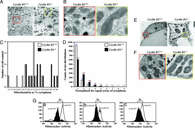
Cyclin D1-deficiency enhances mitochondrial size and function. (A and B) Transmission electron microscopic (TEM) images of hepatocytes from liver tissue of cyclin D1+/+ (Left; red box, enlarged area) and cyclin D1−/− (Right; yellow box, enlarged area) show increased mitochondria size in cyclin D1−/−. Catalase-positive peroxisomes (dark spherical structures) are evident in A. (Magnification: B, ×5,000.) (C and D) Mitochondrial size is increased as a proportion of cellular cytoplasm by stereoscopy in cyclin D1−/− and in cyclin D1+/+ hepatocytes. (E) TEM of mammary gland adipocyte with enlarged view (F) revealing increased mitochondria size. (G) Mitochondrial activity in hepatocytes (Ga) and bone marrow macrophages (Gb) and MEFs (Gc) derived from either cyclin D1+/+ or cyclin D1−/− assessed by using MitoTracker (Deep Red, 50 nM).
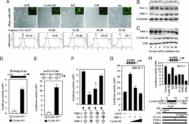
NRF-1 activity is enhanced in cyclin D1-deficient cells. (A) Vectors encoding cyclin D1 and mutants in the vector MSCV-IRES-GFP were used to transduce cyclin D1−/− MEFs for 1 week. Cells were stained with Mitotracker Red CMX Ros for 30 min. The GFP-positive cells were sorted, and MitoTracker fluorescence and cell cycle were analyzed by flow cytometry. The cyclin D1 K112 residue is required for inhibition of mitochondrial activity and induction of DNA synthesis. (B and C) MCF-7 cells were starved in DMEM supplemented with 0.2% FBS for 48 h. Cells were harvested at time points as indicated. Total cell lysates were prepared and subjected to Western blotting analysis to detect cyclin D1 and NRF-1 expression. β-tubulin and GDI were included as loading controls. (D–F) Cyclin D1+/+ and cyclin D1−/− 3T3 cells were transfected with promoters driving luciferase reporter genes for wild-type mtTFA, D loop (D), CMV (E), or mtTFA promoter reporter plasmids encoding mutant response elements for NRF-1, Sp-1, or NRF-2 (F). The luciferase activity is mean ± SEM (n ≥ 6). (G) MCF-7 cells were transfected in 24-well plates with 1 μg of a luciferase reporter gene containing four copies of NRF-1 response elements, 0.5 μg of pSG5 vector or pSG5-NRF-1 expression plasmid, and cyclin D1 expression vector. Luciferase activity is normalized to a cotransfected pRL-TK Luc control. (H) Mutant cyclin D1 expression plasmids were compared for NRF-1 repression function. (mean ± SEM; n > 3 separate experiments each performed in quadruplicate).
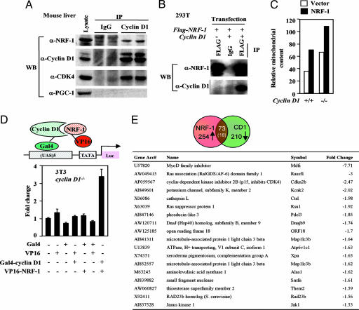
Cyclin D1 interacts with NRF-1. (A) Lysates from mouse liver were subjected to cyclin D1 antibody IP. IP products were resolved on SDS/PAGE with Western blot to NRF-1 or PGC-1. Cyclin D1 associates with NRF-1 but not PGC-1 in vivo. (B) HEK293T cells transfected with FLAG-tagged NRF-1 and cyclin D1. Cell lysates were subjected to either IgG or anti-FLAG antibody IP. IP products were resolved on SDS/PAGE followed by Western blot to either FLAG for NRF-1 or cyclin D1. (C) Vectors encoding NRF-1 in the vector MSCV-IRES-GFP were used to transduce cyclin D1−/− and cyclin D1+/[supi]+ MEFs. Cells were stained with MitoTracker Red CMX Ros for 30 min. The GFP-positive cells were sorted, and MitoTracker fluorescence was analyzed by flow cytometry. (D) The Gal4-cyclin D1 and VP16-NRF-1 fusion constructs were transfected with the pG5luc reporter into cyclin D1−/− 3T3 cells. Firefly luciferase activity was quantitated by using the Dual-Luciferase Reporter Assay System (Promega). Interaction between these two proteins (lane 7) increases luciferase activity over the negative controls. (E) Comparison of cyclin D1-regulated genes determined by microarray analysis (available upon request) with genomewide location analysis of candidate NRF-1 target genes comparing genes found on the HU13K and MGU74 chips.
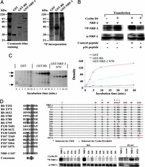
NRF-1 serves as substrate of cyclin D1-dependent kinase. (A) GST-NRF-1 was incubated with immunoprecipitated cyclin D1/Cdk4 kinase complex in the presence of [γ-32P]ATP. (A Left) Coomassie blue staining of input GST protein. (A Right) γ-32P incorporation into GST-NRF-1. GST and GST-RB were negative and positive control for kinase activity. (B Left) HEK293T cells were transfected with FLAG-NRF-1 with cyclin D1 or control empty vector. (B Right) HEK293T cells transfected with FLAG-NRF-1 and cyclin D1 were treated with p16INK4a peptide (20 μM) corresponding to amino acids 84–103 of the human p16INK4a protein (DAAREGFLATLVVLHRAGAR) with a C-terminal 16-aa Penetratin (RQIKIWFQNRRMKWKK) or control peptide (Biosynthesis, Lewisville, TX) (16). NRF-1 phosphorylation was abrogated by p16INK4a peptide. Cells were pulse-labeled with [γ-32P]orthophosphate. NRF-1 protein was precipitated with anti-FLAG M2 antibody and subjected to autoradiography. (C Left) Equal amounts of either the GST-NRF-1 N70 fusion protein or GST protein were incubated with 200 ng of purified cyclin D1/Cdk4 complex and [γ32P]ATP. The arrows indicate the autoradiogram of the phosphorylated fusion protein. (C Right) GST-Rb serves as a positive control. The phosphorylated bands were quantified by densitometry scanning. (D) Alignment of Cdk4 phosphorylation sites for pRb, p107, p130, and NRF-1. HEK293T cells were transfected with expression vectors encoding hemagglutinin or FLAG-tagged NRF-1 and mutants in which a single potential phosphorylation site was restored in all sites mutated version (12xA) or a single point mutant of S47, together with cyclin D1. NRF-1 and mutant protein were precipitated with anti-hemagglutinin antibody and subjected to autoradiography.
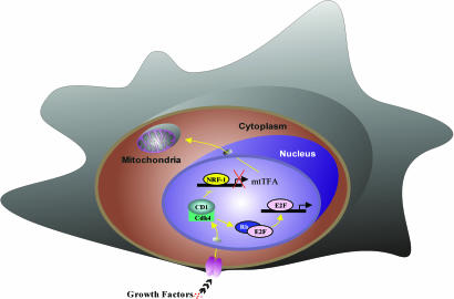
Schematic representation of the mechanism by which cyclin D1 inhibits mitochondrial function. Cyclin D1-dependent kinase phosphorylates and inhibits NRF-1 and, thereby, mtTFA and mitochondrial activity. Additional NRF-1-independent mechanisms regulating mitochondrial activity remain to be defined.
Similar articles
-
Cyclin D1 determines mitochondrial function in vivo.Mol Cell Biol. 2006 Jul;26(14):5449-69. doi: 10.1128/MCB.02074-05. Mol Cell Biol. 2006. PMID: 16809779 Free PMC article.
-
Translokin (Cep57) interacts with cyclin D1 and prevents its nuclear accumulation in quiescent fibroblasts.Traffic. 2011 May;12(5):549-62. doi: 10.1111/j.1600-0854.2011.01176.x. Epub 2011 Mar 15. Traffic. 2011. PMID: 21306487
-
Identification of mutations that disrupt phosphorylation-dependent nuclear export of cyclin D1.Oncogene. 2006 Oct 12;25(47):6291-303. doi: 10.1038/sj.onc.1209644. Epub 2006 May 29. Oncogene. 2006. PMID: 16732330
-
Mitochondrial reactive oxygen species-mediated genomic instability in low-dose irradiated human cells through nuclear retention of cyclin D1.Cell Cycle. 2016 Jun 2;15(11):1410-4. doi: 10.1080/15384101.2016.1170271. Epub 2016 Apr 14. Cell Cycle. 2016. PMID: 27078622 Free PMC article. Review.
-
New roles of cyclin D1.Am J Pathol. 2013 Jul;183(1):3-9. doi: 10.1016/j.ajpath.2013.03.001. Am J Pathol. 2013. PMID: 23790801 Free PMC article. Review.
Cited by
-
An analysis of cyclin D1, cytokeratin 5/6 and cytokeratin 8/18 expression in breast papillomas and papillary carcinomas.Diagn Pathol. 2013 Jan 18;8:8. doi: 10.1186/1746-1596-8-8. Diagn Pathol. 2013. PMID: 23327593 Free PMC article.
-
Cyclin D1 targets hexokinase 2 to control aerobic glycolysis in myeloma cells.Oncogenesis. 2020 Jul 24;9(7):68. doi: 10.1038/s41389-020-00253-3. Oncogenesis. 2020. PMID: 32709889 Free PMC article.
-
Two-way communication between the metabolic and cell cycle machineries: the molecular basis.Cell Cycle. 2015;14(13):2022-32. doi: 10.1080/15384101.2015.1044172. Cell Cycle. 2015. PMID: 26038996 Free PMC article. Review.
-
Small non-coding RNAs govern mammary gland tumorigenesis.J Mammary Gland Biol Neoplasia. 2012 Mar;17(1):59-64. doi: 10.1007/s10911-012-9246-4. Epub 2012 Mar 1. J Mammary Gland Biol Neoplasia. 2012. PMID: 22382486 Free PMC article. Review.
-
CDK2 regulates the NRF1/Ehmt1 axis during meiotic prophase I.J Cell Biol. 2019 Sep 2;218(9):2896-2918. doi: 10.1083/jcb.201903125. Epub 2019 Jul 26. J Cell Biol. 2019. PMID: 31350280 Free PMC article.
References
-
- Shadel G. S., Clayton D. A. Annu. Rev. Biochem. 1997;66:409–435. - PubMed
-
- Larsson N. G., Wang J., Wilhelmsson H., Oldfors A., Rustin P., Lewandoski M., Barsh G. S., Clayton D. A. Nat. Genet. 1998;18:231–236. - PubMed
-
- Evans M. J., Scarpulla R. C. Genes Dev. 1990;4:1023–1034. - PubMed
-
- Scarpulla R. C. Biochim. Biophys. Acta. 2002;1576:1–14. - PubMed
Publication types
MeSH terms
Substances
Grants and funding
LinkOut - more resources
Full Text Sources
Molecular Biology Databases
Research Materials
