. 2006 Aug 1;103(31):11766-71.
doi: 10.1073/pnas.0601182103. Epub 2006 Jul 24.

Adaptive response of Yersinia pestis to extracellular effectors of innate immunity during bubonic plague

Affiliations

Adaptive response of Yersinia pestis to extracellular effectors of innate immunity during bubonic plague

Florent Sebbane et al. Proc Natl Acad Sci U S A. .

Abstract

Yersinia pestis causes bubonic plague, characterized by an enlarged, painful lymph node, termed a bubo, that develops after bacterial dissemination from a fleabite site. In susceptible animals, the bacteria rapidly escape containment in the lymph node, spread systemically through the blood, and produce fatal sepsis. The fulminant progression of disease has been largely ascribed to the ability of Y. pestis to avoid phagocytosis and exposure to antimicrobial effectors of innate immunity. In vivo microarray analysis of Y. pestis gene expression, however, revealed an adaptive response to nitric oxide (NO)-derived reactive nitrogen species and to iron limitation in the extracellular environment of the bubo. Polymorphonuclear neutrophils recruited to the infected lymph node expressed abundant inducible NO synthase, and several Y. pestis homologs of genes involved in the protective response to reactive nitrogen species were up-regulated in the bubo. Mutation of one of these genes, which encodes the Hmp flavohemoglobin that detoxifies NO, attenuated virulence. Thus, the ability of Y. pestis to destroy immune cells and remain extracellular in the bubo appears to limit exposure to some but not all innate immune effectors. High NO levels induced during plague may also influence the developing adaptive immune response and contribute to septic shock.


Conflict of interest statement

Conflict of interest statement: No conflicts declared.

Figures

Fig. 1.
Fig. 1.
Principal component analysis of microarray data from six biological replicate samples for each of the in vitro and in vivo conditions. The first two principal components show the separation of the bubo-specific expression profile from the expression profiles in exponential and stationary-phase cultures grown at 21°C and 37°C. Ellipses encompass exponential, stationary, and rat samples.
Fig. 2.
Fig. 2.
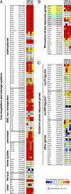
Differential expression in the bubo of genes implicated in the response to iron limitation (A) (9), nitrosative stress (B) (–20), and oxidative stress (C) (22, 39). The mean fold increase or decrease in individual gene expression in the bubo compared with exponential (E) and stationary (S) phase cultures at 21°C and 37°C is indicated by the color scale bar. For the nitrosative stress gene list (B), genes specifically required for resistance to NO and GSNO or to peroxynitrite are highlighted in yellow and green, respectively. Genes highlighted in gray, along with hmp and ytfE, are predicted to be up-regulated in response to RNS by repression of the NO-sensitive negative regulator encoded by nsrR (17, 18); the other genes were shown to be up-regulated at least 2-fold in response to NO or GSNO stress in three different in vitro microarray studies (–15). The YPO and Y numbers refer to the Y. pestis CO92 and KIM genome annotations, respectively (10, 11). ∗, not annotated in Y. pestis CO92; a, pseudogene; b, the Y. pestis response to superoxide may not occur via SoxRS regulation because no soxRS homologs were identified in the genome (10, 11).
Fig. 3.
Fig. 3.
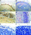
iNOS expression by PMNs in the bubo. Shown are sections of a bubo (AD) or uninfected lymph node (E and F) stained to detect iNOS (A, C, and E) or PMNs (B, D, and F; yellow arrowheads). PMNs producing iNOS (dark brown) are indicated by red arrowheads. Masses of extracellular bacteria adjacent to PMNs are indicated by blue arrowheads. (Magnification: A and B, ×100; D, ×400; C, E, and F, ×600.)
Fig. 4.
Fig. 4.
Decreased resistance of Y. pestis to RNS attenuates virulence more than decreased resistance to ROS. (A and B) Susceptibility of Y. pestis wild type (black bars), Δhmp (gray bars), and ΔsodA (white bars) to RNS and ROS in vitro (A) and to intracellular killing by macrophages (B). The mean and standard error of three experiments are shown. (C and D) Incidence of plague in rats injected intradermally with ≈150 Y. pestis wild type (black circles), Δhmp (gray circles), or ΔsodA (white circles).

Similar articles

Cited by

References

    1. Perry R. D., Fetherston J. D. Clin. Microbiol. Rev. 1997;10:35–66. - PMC - PubMed
    1. Sebbane F., Gardner D., Long D., Gowen B. B., Hinnebusch B. J. Am. J. Pathol. 2005;166:1427–1439. - PMC - PubMed
    1. Cornelis G. R., Boland A., Boyd A. P., Geuijen C., Iriarte M., Neyt C., Sory M. P., Stainier I. Microbiol. Mol. Biol. Rev. 1998;62:1315–1352. - PMC - PubMed
    1. Navarro L., Alto N. M., Dixon J. E. Curr. Opin. Microbiol. 2005;8:21–27. - PubMed
    1. Une T., Brubaker R. R. Infect. Immun. 1984;43:895–900. - PMC - PubMed

Publication types

MeSH terms

Associated data