

Structural basis for unique mechanisms of folding and hemoglobin binding by a malarial protease
- PMID: 16864794
- PMCID: PMC1544199
- DOI: 10.1073/pnas.0600489103
Structural basis for unique mechanisms of folding and hemoglobin binding by a malarial protease
Abstract
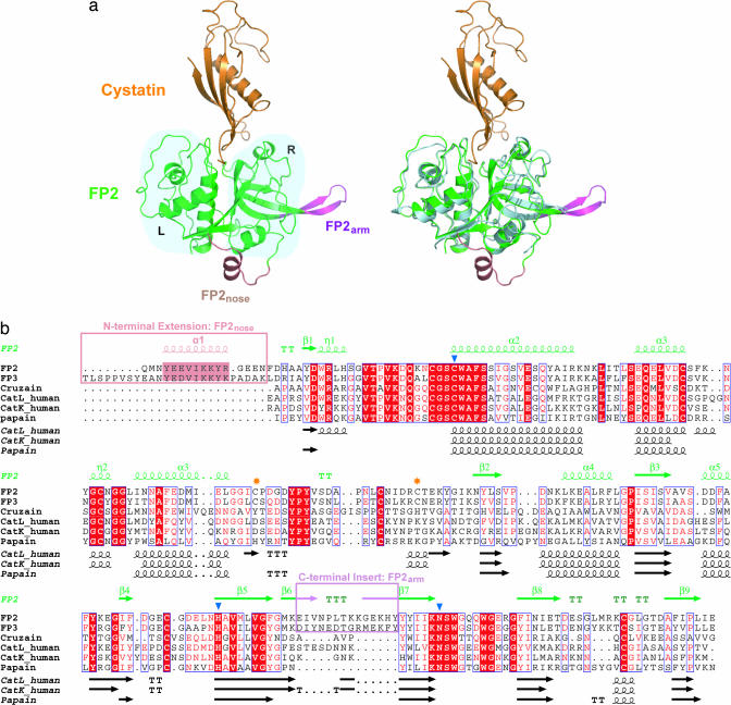
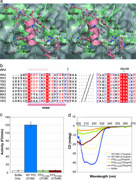
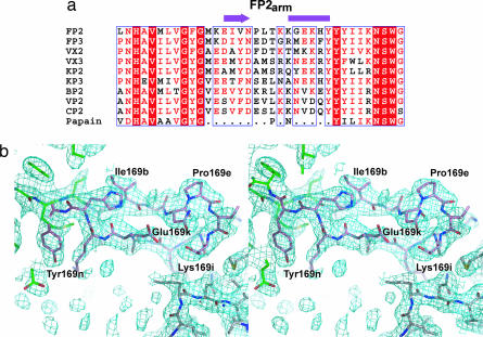
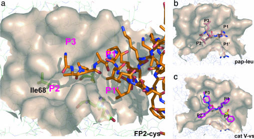
Falcipain-2 (FP2), the major cysteine protease of the human malaria parasite Plasmodium falciparum, is a hemoglobinase and promising drug target. Here we report the crystal structure of FP2 in complex with a protease inhibitor, cystatin. The FP2 structure reveals two previously undescribed cysteine protease structural motifs, designated FP2(nose) and FP2(arm), in addition to details of the active site that will help focus inhibitor design. Unlike most cysteine proteases, FP2 does not require a prodomain but only the short FP2(nose) motif to correctly fold and gain catalytic activity. Our structure and mutagenesis data suggest a molecular basis for this unique mechanism by highlighting the functional role of two Tyr within FP2(nose) and a conserved Glu outside this motif. The FP2(arm) motif is required for hemoglobinase activity. The structure reveals topographic features and a negative charge cluster surrounding FP2(arm) that suggest it may serve as an exo-site for hemoglobin binding. Motifs similar to FP2(nose) and FP2(arm) are found only in related plasmodial proteases, suggesting that they confer malaria-specific functions.
Conflict of interest statement
Conflict of interest statement: No conflicts declared.
Figures
Similar articles
-
The Plasmodium falciparum cysteine protease falcipain-2 captures its substrate, hemoglobin, via a unique motif.Proc Natl Acad Sci U S A. 2005 Jun 28;102(26):9138-43. doi: 10.1073/pnas.0502368102. Epub 2005 Jun 17. Proc Natl Acad Sci U S A. 2005. PMID: 15964982 Free PMC article.
-
Independent intramolecular mediators of folding, activity, and inhibition for the Plasmodium falciparum cysteine protease falcipain-2.J Biol Chem. 2004 Jan 30;279(5):3484-91. doi: 10.1074/jbc.M310536200. Epub 2003 Nov 18. J Biol Chem. 2004. PMID: 14625277
-
New insights of falcipain 2 structure from Plasmodium falciparum 3D7 strain.Biochem Biophys Res Commun. 2022 Jan 29;590:145-151. doi: 10.1016/j.bbrc.2021.12.080. Epub 2021 Dec 23. Biochem Biophys Res Commun. 2022. PMID: 34974303
-
Falcipains and other cysteine proteases of malaria parasites.Adv Exp Med Biol. 2011;712:30-48. doi: 10.1007/978-1-4419-8414-2_3. Adv Exp Med Biol. 2011. PMID: 21660657 Review.
-
Cysteine proteases of malaria parasites.Int J Parasitol. 2004 Dec;34(13-14):1489-99. doi: 10.1016/j.ijpara.2004.10.003. Int J Parasitol. 2004. PMID: 15582526 Review.
Cited by
-
The Potential of Secondary Metabolites from Plants as Drugs or Leads against Protozoan Neglected Diseases-Part III: In-Silico Molecular Docking Investigations.Molecules. 2016 Oct 19;21(10):1389. doi: 10.3390/molecules21101389. Molecules. 2016. PMID: 27775577 Free PMC article. Review.
-
Cross-talk between malarial cysteine proteases and falstatin: the BC loop as a hot-spot target.PLoS One. 2014 Apr 3;9(4):e93008. doi: 10.1371/journal.pone.0093008. eCollection 2014. PLoS One. 2014. PMID: 24699522 Free PMC article.
-
Hemoglobin cleavage site-specificity of the Plasmodium falciparum cysteine proteases falcipain-2 and falcipain-3.PLoS One. 2009;4(4):e5156. doi: 10.1371/journal.pone.0005156. Epub 2009 Apr 9. PLoS One. 2009. PMID: 19357776 Free PMC article.
-
Structures of falcipain-2 and falcipain-3 bound to small molecule inhibitors: implications for substrate specificity.J Med Chem. 2009 Feb 12;52(3):852-7. doi: 10.1021/jm8013663. J Med Chem. 2009. PMID: 19128015 Free PMC article.
-
The complex of Plasmodium falciparum falcipain-2 protease with an (E)-chalcone-based inhibitor highlights a novel, small, molecule-binding site.Malar J. 2019 Dec 2;18(1):388. doi: 10.1186/s12936-019-3043-0. Malar J. 2019. PMID: 31791339 Free PMC article.
References
Publication types
MeSH terms
Substances
Associated data
- Actions
Grants and funding
LinkOut - more resources
Full Text Sources
Molecular Biology Databases
