. 2006 Aug 1;103(31):11790-5.
doi: 10.1073/pnas.0604400103. Epub 2006 Jul 24.

Microsomal prostaglandin E synthase-1 is a critical factor of stroke-reperfusion injury

Affiliations

Microsomal prostaglandin E synthase-1 is a critical factor of stroke-reperfusion injury

Yuri Ikeda-Matsuo et al. Proc Natl Acad Sci U S A. .

Abstract

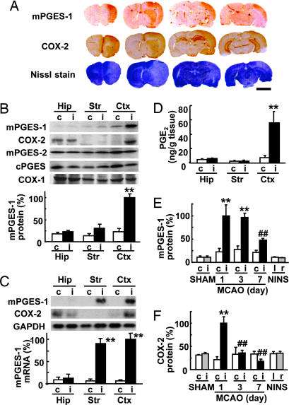
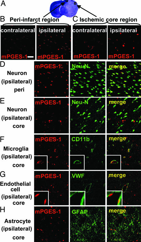
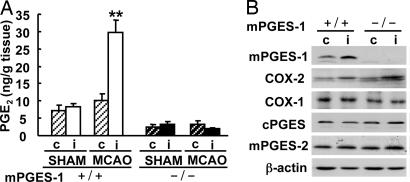
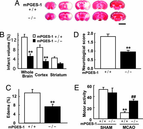
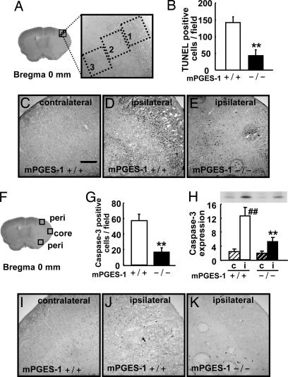
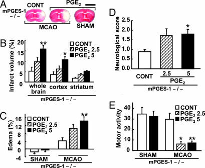
Although augmented prostaglandin E(2) (PGE(2)) synthesis and accumulation have been demonstrated in the lesion sites of rodent transient focal ischemia models, the role of PGE(2) in neuronal survival has been controversial, showing both protective and toxic effects. Here we demonstrate the induction of microsomal PGE synthase 1 (mPGES-1), an inducible terminal enzyme for PGE(2) synthesis, in neurons, microglia, and endothelial cells in the cerebral cortex after transient focal ischemia. In mPGES-1 knockout (KO) mice, in which the postischemic PGE(2) production in the cortex was completely absent, the infarction, edema, apoptotic cell death, and caspase-3 activation in the cortex after ischemia were all reduced compared with those in wild-type (WT) mice. Furthermore, the behavioral neurological dysfunctions observed after ischemia in WT mice were significantly ameliorated in KO mice. The ameliorated symptoms observed in KO mice after ischemia were reversed to almost the same severity as WT mice by intracerebroventricular injection of PGE(2) into KO mice. Our observations suggest that mPGES-1 may be a critical determinant of postischemic neurological dysfunctions and a valuable therapeutic target for treatment of human stroke.


Conflict of interest statement

Conflict of interest statement: No conflicts declared.

Figures

Fig. 1.
Fig. 1.
mPGES-1 induction in the rat brain after transient ischemia. (A) Immunostaining for mPGES-1 and COX-2 and Nissle staining of a coronal brain slice 24 h after ischemia. Representative data from six animals are presented. (Scale bar, 5 mm.) (B) (Upper) Western blot analysis of the expression of mPGES-1, COX-2, mPGES-2, cPGES, and COX-1 in the hippocampus (Hip), striatum (Str), and cortex (Ctx) of the ipsilateral (i) or contralateral (c) hemisphere 24 h after ischemia. (Lower) Quantitated data from immunoblotting with mPGES-1 antibody were scaled to a percentage of the maximal response. (C) (Upper) Northern blot analysis of the expression of mPGES-1 and COX-2 in the brain tissue 6 h after ischemia. GAPDH signals were used as loading controls. (Lower) Quantitated data from blotting with mPGES-1 probe were normalized to GAPDH and scaled to a percentage of the maximal response. (D) The amount of PGE2 in the brain tissue 24 h after ischemia was measured by using an enzyme immunoassay kit. (E and F) Time course of mPGES-1 (E) and COX-2 (F) protein expression in the cortex of MCAO animals, sham-operated animals killed 24 h after surgery (SHAM), and animals without surgery and ischemia (no-ischemia, no-surgery; NINS). Quantitated data from immunoblotting with mPGES-1 and COX-2 antibody were scaled to a percentage of the maximal response. n = 4 animals per group; ∗∗, P < 0.01 vs. the contralateral tissue; ##, P < 0.01; and #, P < 0.05 vs. the ipsilateral cortex (day 1).
Fig. 2.
Fig. 2.
Induction of mPGES-1 in neurons, microglia, and endothelial cells in the cortex after ischemia. (A) The predesignated areas in the peri-infarct and ischemic core regions of the postischemic cortex for microscopic examination. (B and C) The immunostaining of mPGES-1 in the peri-infarct region (B) and ischemic core region (C) of ipsilateral postischemic cortex and contralateral cortex. (DH) The double-immunostaining of mPGES-1 (red) and cell-type-specific marker proteins (green) in the peri-infarct (D) and core (EH) regions of the cortex. Neurons (D and E), microglia (F), endothelial cells (G), and astrocytes (H) were recognized by antibodies for Neu-N, CD11b, von Willebrand factor (VWF), and GFAP, respectively. Insets show high magnification of each staining. The photographs shown are representative examples from three separate experiments. (Scale bars: main images, 40 μm; Insets, 10 μm.)
Fig. 3.
Fig. 3.
mPGES-1 is an essential component for PGE2 production in the postischemic cortex. (A) The production of PGE2 in the ipsilateral (i) or contralateral (c) cortex of mPGES-1 KO (−/−) and WT (+/+) mice 24 h after MCAO and sham operation. n = 8 animals per group; ∗∗, P < 0.01 vs. another sample. (B) Western blot analyses for mPGES-1, COX-2, COX-1, cPGES, and mPGES-2 in the cortex 24 h after transient ischemia. Representative immunoblots from three separate experiments are presented.
Fig. 4.
Fig. 4.
Deletion of the mPGES-1 resulted in marked amelioration of the infarction, edema, and behavioral neurological dysfunctions observed after ischemia. (A) Representative 2,3,5-triphenyltetrazolium chloride (TTC)-stained coronal sections at −2, 0, +2, +4, and +8 mm from the bregma of the mPGES-1 KO (−/−) and WT (+/+) mouse. (Scale bar: 5 mm.) (B) The volume of infarcted brain tissue 24 h after ischemia was estimated and expressed as a percentage of the corrected tissue volume. n = 10 animals per group; ∗∗, P < 0.01 vs. WT mice. (C) The corrected edema percentage in the mPGES-1 KO (−/−) and WT (+/+) mice. n = 10 animals per group; ∗∗, P < 0.01 vs. WT mice. (D) Improvement of neurological dysfunction in mPGES-1 KO mice. The neurological score was measured 24 h after ischemia. n = 21–22 animals per group; ∗∗, P < 0.01 vs. WT mice. (E) The motor activity of MCAO or sham-operated (SHAM) mice. n = 9–10 animals for SHAM and n = 19–20 animals for MCAO; ∗∗, P < 0.01 vs. SHAM WT mice; ##, P < 0.01 vs. MCAO-treated WT mice.
Fig. 5.
Fig. 5.
Deletion of the mPGES-1 gene resulted in a marked decrease in the apoptotic neuronal death in the postischemic cortex. (A) Three predesignated areas in the penumbra of the postischemic cortex for counting TUNEL-positive cells. (B) The average number of TUNEL-positive cells per unit area (A) of mPGES-1 KO (−/−) and WT (+/+) mice. n = 7 animals per group; ∗∗, P < 0.01 vs. WT mice. (CE) Representative results of TUNEL staining in the contralateral or ipsilateral cortex of the mPGES-1 KO and WT mouse. (Scale bar: 100 μm.) (F) Three predesignated areas in peri-infarct (peri) and core regions for counting caspase-3-positive cells in the cortex. (G) The average number of caspase-3-positive cells per unit area (F) after transient ischemia. n = 7 animals per group; ∗∗, P < 0.01 vs. WT mice. (H) The tissue lysates from the contralateral (c) or ipsilateral (i) cortex were subjected to Western blot analysis for caspase-3. The densities of immunoblots were measured. n = 5 animals per group; ##, P < 0.01 vs. contralateral cortex. (IK) Representative caspase-3 immunostaining in the cortex.
Fig. 6.
Fig. 6.
i.c.v. injection of PGE2 exaggerated postischemic symptoms in mPGES-1 KO mice. (A) Representative 2,3,5-triphenyltetrazolium chloride (TTC)-stained coronal sections (3 mm from the prefrontal) from PGE2-injected (5 ng) or vehicle-injected (CONT) mice brain 24 h after ischemia (MCAO) or sham operation. (Scale bar: 5 mm.) (B and C) The corrected volume of infarcted brain tissue (B) and the corrected edema percentage (C) were measured from PGE2-injected (2.5 or 5 ng) or vehicle-injected (CONT) injected mice brain 24 h after ischemia (MCAO) or sham operation and expressed as a percentage of the total volume. (D and E) The neurological score (D) and the motor activity (E) were measured 24 h after ischemia (MCAO) or sham operation. For BE, n = 7–10; ∗∗, P < 0.01; ∗, P < 0.05 vs. CONT.

Similar articles

Cited by

References

    1. Lyden P. D., Grotta J. C., Levine S. R., Marler J. R., Frankel M. R., Brott T. G. Neurology. 1997;49:14–20. - PubMed
    1. Lyden P. D. Prog. Cardiovasc. Dis. 1999;42:175–183. - PubMed
    1. Koistinaho J., Hokfelt T. NeuroReport. 1997;8:i–viii. - PubMed
    1. Dirnagl U., Iadecola C., Moskowitz M. A. Trends Neurosci. 1999;22:391–397. - PubMed
    1. Barone F. C., Feuerstein G. Z. J. Cereb. Blood Flow Metab. 1999;19:819–834. - PubMed

Publication types

MeSH terms