. 2006 Aug 2;128(30):9809-12.
doi: 10.1021/ja0615425.

Investigating a quadruplex-ligand interaction by unfolding kinetics

Affiliations

Investigating a quadruplex-ligand interaction by unfolding kinetics

Jeremy J Green et al. J Am Chem Soc. .

Abstract

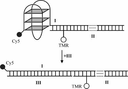
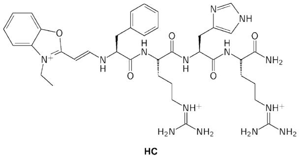
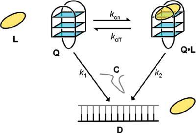
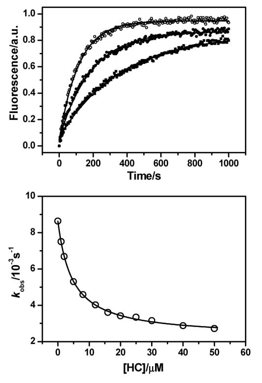
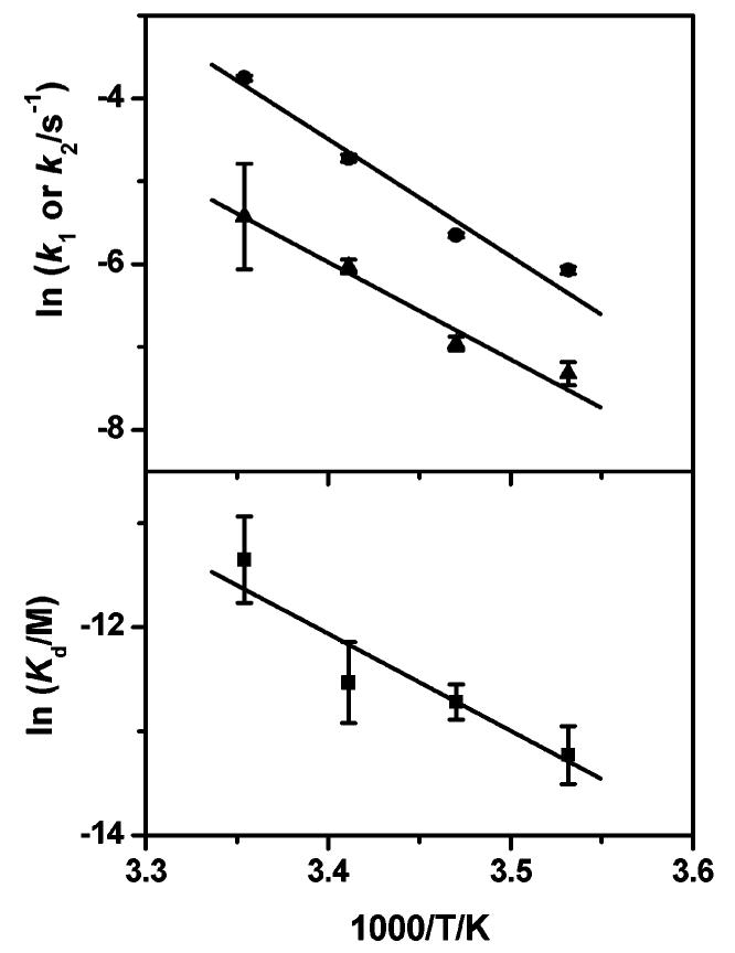
We have investigated the interaction of the intramolecular human telomeric DNA G-quadruplex with a hemicyanine-peptide ligand, by studying the rate of quadruplex opening with a complementary DNA oligonucleotide. By employing a minimal kinetic model, the relationship between the observed rate of quadruplex opening and the ligand concentration has enabled estimation of the dissociation constant. A van't Hoff analysis revealed the enthalpy and entropy changes of binding to be -77 +/- 22 kJ mol(-1) and -163 +/- 75 J mol(-1) K(-1), respectively. Arrhenius analyses of the rate constants of opening free and bound quadruplex gave activation energies of 118 +/- 2 and 98 +/- 10 kJ mol(-1), respectively. These results indicate that the presence of the ligand has only a small effect on the activation energy, suggesting that the unbinding of the ligand occurs after the transition state for quadruplex unfolding.


Figures

Figure 1
Figure 1
FRET-based quadruplex system used in this study. Roman numerals represent the labels given to strands (see Materials and Methods), and TMR and Cy5 are the fluorophores that make up a FRET pair. Quadruplex opening is followed using the change in FRET efficiency between the TMR and Cy5.
Figure 2
Figure 2
Structure of the hemicyanine tetrapeptide conjugate HC.
Figure 3
Figure 3
Scheme used to derive a kinetic model of the opening of quadruplex Q in the presence of ligand L and complementary strand C to give duplex D.
Figure 4
Figure 4
Top: the increase of TMR fluorescence monitored at 580 nm as the quadruplex formed by I:II is opened by III in 10 mM Tris·HCl (pH 7.4) and 100 mM NaCl at 20 °C. Data from runs performed in the presence of ligand HC at concentrations 0 (○), 5 μM (●), and 50 μM (■) are shown, along with single-exponential fits (solid lines). In each series the initial fluorescence intensity has been subtracted from each data point. The rate constants of opening, kobs, were found to be 0.0086, 0.0053, and 0.0027 s−1 at 0, 5, and 50 μM HC, respectively. Bottom: the variation of observed rate constant with concentration of HC for the hybridization of I:II to III at 20 °C in 10 mM Tris·HCl (pH 7.4) and 100 mM NaCl. The data are shown as open circles and fits to eq 4 as a solid line.
Figure 5
Figure 5
Top: Arrhenius plots of the opening rates of I:II (k1, ●) and HC·I:II (k2, ▲), respectively. Bottom: van't Hoff plot of the dissociation constant (Kd, ■) of HC to I:II. Activation energies for k1 and k2 obtained from the fits are 118 ± 2 and 98 ± 10 kJ mol−1, respectively, and the thermodynamic parameters found are ΔH = −77 ± 22 kJ mol−1 and ΔS = −163 ± 75 J mol−1 K−1.

References

    1. Sen D, Gilbert W. Nature. 1988;334:364–366. - PubMed
    1. Gellert M, Lipsett MN, Davies DR. Proc. Natl. Acad. Sci. U.S.A. 1962;48:2013–2018. - PMC - PubMed
    1. Van Dyke MW, Nelson LD, Weilbaecher RG, Mehta DV. J. Biol. Chem. 2004;279:24323–24333. - PubMed
    1. Tolstonog GV, Li G, Shoeman RL, Traub P. DNA Cell Biol. 2005;24:85–110. - PubMed
    1. Zaug AJ, Podell ER, Cech TR. Proc. Natl. Acad. Sci. U.S.A. 2005;102:10864–10869. - PMC - PubMed

Publication types