Comparative Study
. 2006 Nov 15;108(10):3313-20.
doi: 10.1182/blood-2006-03-006908. Epub 2006 Jul 25.

Correction of the disease phenotype in canine leukocyte adhesion deficiency using ex vivo hematopoietic stem cell gene therapy

Affiliations
Comparative Study

Correction of the disease phenotype in canine leukocyte adhesion deficiency using ex vivo hematopoietic stem cell gene therapy

Thomas R Bauer Jr et al. Blood. .

Abstract

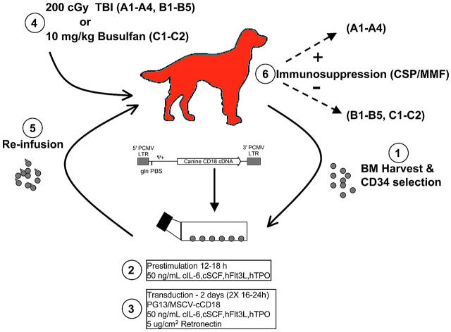
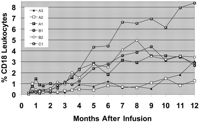
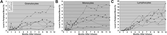
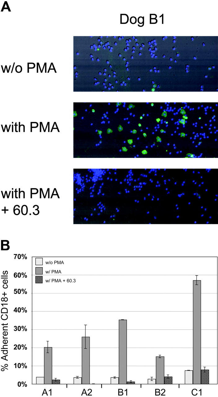
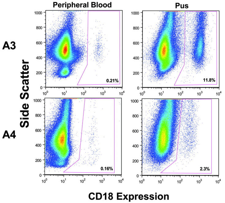
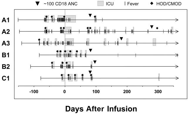
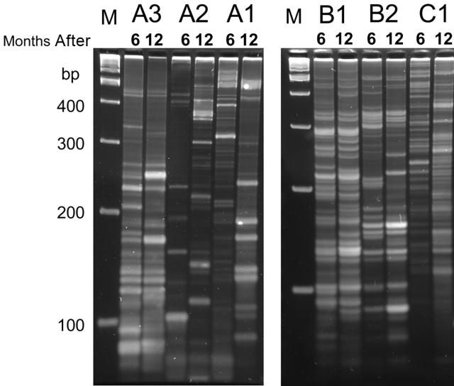
Canine leukocyte adhesion deficiency (CLAD) represents the canine counter-part of the human disease leukocyte adhesion deficiency (LAD). Defects in the leukocyte integrin CD18 adhesion molecule in both CLAD and LAD lead to recurrent, life-threatening bacterial infections. We evaluated ex vivo retroviral-mediated gene therapy in CLAD using 2 nonmyeloablative conditioning regimens--200 cGy total body irradiation (TBI) or 10 mg/kg busulfan--with or without posttransplantation immunosuppression. In 6 of 11 treated CLAD dogs, therapeutic levels of CD18(+) leukocytes were achieved. Conditioning with either TBI or busulfan allowed long-term engraftment, and immunosuppression was not required for efficacy. The percentage of CD18(+) leukocytes in the peripheral blood progressively increased over 6 to 8 months after infusion to levels ranging from 1.26% to 8.37% at 1-year follow-up in the 6 dogs. These levels resulted in reversal or moderation of the severe CLAD phenotype. Linear amplification-mediated polymerase chain reaction assays indicated polyclonality of insertion sites. These results describe ex vivo hematopoietic stem cell gene transfer in a disease-specific, large animal model using 2 clinically applicable conditioning regimens, and they provide support for the use of nonmyeloablative conditioning regimens in preclinical protocols of retroviral-mediated gene transfer for nonmalignant hematopoietic diseases such as LAD.


Figures

Figure 1.
Figure 1.
Diagram of experimental design. Bone marrow (BM) was harvested from CLAD dogs and enriched for CD34+ cells. CD34+ cells were used immediately, or cryopreserved and thawed 2 to 3 weeks later. After transduction, cells were harvested, washed, and reinfused into CLAD pups that had received conditioning with either 200 cGy TBI on day 0 or –1 (n = 9), or 10 mg/kg busulfan intravenously on day –2(n = 2). Four dogs (A1-A4) received posttransplantation immunosuppression with CSP starting on day –1 and continuing until day +65 (35 days at 30 mg/kg, 30 days at 15 mg/kg), along with MMF from day 0 to day +28 (at 20 mg/kg). Seven dogs received no posttransplantation immunosuppression. LTR, retroviral long terminal repeat; Ψ+, extended packaging signal; gln tRNA PBS, glutamine tRNA primer binding site; PCMV, PCC4-cell passaged myeloproliferative sarcoma virus.
Figure 2.
Figure 2.
Flow cytometric analysis of CD18+ leukocytes in the peripheral blood following infusion of gene-corrected cells. Peripheral-blood leukocytes from the 6 CLAD dogs surviving long-term were isolated, stained with an anti-CD18 antibody, and examined by flow cytometry. The percentage of CD18+ leukocytes was determined in comparison to isotype stained controls. Prior to the infusion of the gene-corrected CD34+ cells, there were no CD18+ leukocytes (not different from isotype control staining) in the peripheral blood. The CD18+ leukocyte percentages over time in the peripheral blood are shown.
Figure 3.
Figure 3.
Assessment of CD18+ peripheral-blood leukocyte subsets in the 6 treated CLAD dogs with 1-year follow-up. The contribution of each subset was assessed in each of the 6 long-term treated dogs by flow cytometric analysis of CD18 expression on (A) granulocytes, (B) CD14+ monocytes, and (C) lymphocytes. Each subset was analyzed using a specific subset antibody, or was determined by the absence of CD14 staining and side scatter (SSC) profile (high SSC, granulocytes; low SSC, lymphocytes), along with an anti-CD18 antibody.
Figure 4.
Figure 4.
In vitro detection of adhesion by activated CD18+ leukocytes. Peripheral-blood leukocytes from dogs A1, A2, B1, B2, and C1 were added to fibrinogen-coated wells with or without stimulation by PMA or blocking by the anti-CD18 antibody 60.3 antibody. (A) Photo-multiplier tube (PMT) reconstructed images of field images (500 μm × 192 μm) of representative areas of the wells from dog B1 using scatter, CD18 green fluorescence, and blue fluorescence for Hoechst-stained nucleated cells. Without PMA stimulation, few of the adherent leukocytes are CD18+ (without PMA; top panel). Activated CD18+ leukocytes are present following PMA stimulation (with PMA; middle panel). Blocking of PMA-stimulated CD18+ leukocytes was accomplished using the anti-CD18 monoclonal antibody 60.3 (with PMA + 60.3; bottom panel). (B) Cell adherence was assessed using total peripheral-blood leukocytes from 5 treated CLAD dogs. The number of adherent leukocytes that are CD18+, divided by the total number of adherent CD18+ and CD18 leukocytes present, indicates the percentage of adherent leukocytes that are CD18+. The percentage of nonspecific adherence is influenced by washing stringency, hence the ratio of the stimulated-to-nonstimulated percentages is a more accurate indicator of preferential adhesion than the total number of cells binding. The average of 2 experiments is shown with the range indicated.
Figure 5.
Figure 5.
Selective migration of CD18+ leukocytes in vivo. Analysis of CD18+ leukocytes in the peripheral-blood and pus collected at the same time in 2 CLAD dogs after infusion of CD18+ gene-corrected cells demonstrated in vivo migration of CD18+ leukocytes. Leukocytes were isolated from the peripheral blood and pus from infected tissue (dog A3, week 3; dog A4, week 9) and stained for CD18. Dotplots represent CD18 expression (x-axis) versus side-scatter (y-axis). Percentages of CD18+ leukocytes are shown at the lower right corner.
Figure 6.
Figure 6.
Clinical course of 6 long-term surviving CLAD dogs following gene therapy. The clinical course of the 6 long-term CLAD survivors treated with gene therapy is shown prior to and following infusion of gene-corrected cells. The line and arrow indicate the period of follow-up for each dog. Episodes of fever (≥ 102.4°F) necessitating a more aggressive treatment (increased antibiotics/fluids) are shown by the vertical lines. The shaded boxes indicate periods during which the animal was in intensive care. The appearance of an episode of HOD (lameness/joint swelling/leg pain) or CMO (bony jaw/cranial swelling/soreness) is indicated by the closed diamonds. The day of infusion (day 0) is indicated. Attainment of a CD18+ ANC level of 100 ± 5 cells/μL is indicated by the inverted pyramid. Time in days in relationship to the infusion is shown on the x-axis in the panel.
Figure 7.
Figure 7.
Clonality analysis by linear amplification–mediated PCR (LAM-PCR). Genomic DNA was isolated from peripheral-blood leukocytes of 6 dogs at 2 time points (6 and 12 months after infusion) and subjected to LAM-PCR to examine clonality of retroviral insertions. The presence of a band likely indicates a unique insertion site. Samples were electrophoresed on a Spreadex gel and photographed by a digital camera. M indicates marker; bp, base pairs.

Similar articles

Cited by

References

    1. Anderson DC, Schmalsteig FC, Finegold MJ, et al. The severe and moderate phenotypes of heritable Mac-1, LFA-1 deficiency: their quantitative definition and relation to leukocyte dysfunction and clinical features. J Infect Dis. 1985;152: 668-689. - PubMed
    1. Kishimoto TK, Hollander N, Roberts TM, Anderson DC, Springer TA. Heterogenous mutations in the β subunit common to the LFA-1, Mac-1, and p150,95 glycoproteins cause leukocyte adhesion deficiency. Cell. 1987;50: 193-202. - PubMed
    1. Thomas C, Le Deist F, Cavazzana-Calvo M, et al. Results of allogeneic bone marrow transplantation in patients with leukocyte adhesion deficiency. Blood. 1995;86: 1629-1635. - PubMed
    1. Bauer TR Jr, Gu YC, Tuschong LM, et al. Nonmyeloablative hematopoietic stem cell transplantation corrects the disease phenotype in the canine model of leukocyte adhesion deficiency. Exp Hematol. 2005;33: 706-712. - PubMed
    1. Creevy KE, Bauer Jr TR, Tuschong LM, et al. Mixed chimeric hematopoietic stem cell transplant reverses the disease phenotype in canine leukocyte adhesion deficiency. Vet Immunol Immunopathol. 2003;95: 113-121. - PubMed

Publication types