. 2006 Aug 21;95(4):532-40.
doi: 10.1038/sj.bjc.6603276. Epub 2006 Jul 25.

CEACAM6 gene expression in intrahepatic cholangiocarcinoma

Affiliations

CEACAM6 gene expression in intrahepatic cholangiocarcinoma

K Ieta et al. Br J Cancer. .

Abstract

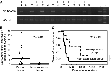
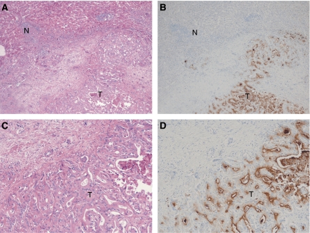
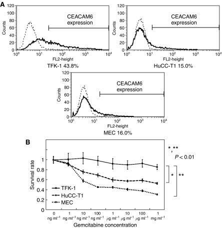
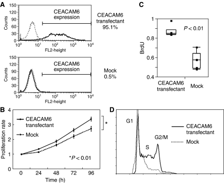
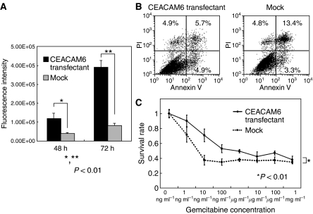
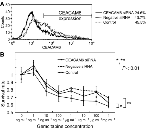
The purpose of this study was to investigate the clinicopathological and biological significance of human carcinoembryonic antigen-related cell adhesion molecule 6 (CEACAM6) gene expression in human intrahepatic cholangiocarcinoma. CEACAM6 is reported to be involved in human malignancies. However, in cholangiocarcinoma expression of CEACAM6 and its clinicopathological significance have not been investigated. CEACAM6 expression status was determined and analysed with respect to various clinicopathological parameters in 23 intrahepatic cholangiocarcinomas. Additionally, we investigated effects of CEACAM6 gene in the cholangiocarcinoma cell lines. CEACAM6 gene expression in cancer tissues was higher than in noncancerous tissues in 16 of the 23 cases; however, it was not statistically significant. The tumours with elevated CEACAM6 expression showed a tendency to be associated with lymphatic invasion and stage of the disease. Interestingly, patients with high CEACAM6 expression showed a significantly poorer disease-free survival rate than those with low CEACAM6 expression. We demonstrated that CEACAM6-transfected cells were more proliferative, more invasive and more chemoresistant to gemcitabine compared to mock-transfected cells. Furthermore, CEACAM6 gene silencing by CEACAM6-specific siRNA resulted in higher chemosensitivity to gemcitabine. CEACAM6 is a potential prognostic indicator and potential chemoresistant marker to gemcitabine for patients with intrahepatic cholangiocarcinoma.


Figures

Figure 1
Figure 1
(A) Expression of CEACAM6 by RT–PCR in representative intrahepatic cholangiocarcinoma patient tissues (T: cancer tissue, N: noncancerous tissue, n: negative control, p: positive control, m: indicates marker). (B) CEACAM6 mRNA expression in cancer and noncancerous tissue with intrahepatic cholangiocarcinoma patients by real-time PCR (n=23). Horizontal lines indicate means. (C) Kaplan–Meier disease-free survival curves in patients with intrahepatic cholangiocarcinoma according to the level of CEACAM6 mRNA expression. The recurrence rate for patients in the high expression group was significantly higher than that for patients in the low expression group (P<0.05). High expression group (n=10): CEACAM6/GAPDH ⩾2, Low expression group (n=13): CEACAM6/GAPDH <2.
Figure 2
Figure 2
Immunohistochemistry with CEACAM6 antibody in intrahepatic cholangiocarcinoma. The majority of stain was observed in cancer cells. (A) original magnification × 40, haematoxylin and eosin stain, (B) original magnification × 40, CEACAM6 stain, (C) original magnification × 100, haematoxylin and eosin stain, (D) original magnification × 100, CEACAM6 stain, T: cancer tissue, N: noncancerous tissue.
Figure 3
Figure 3
(A) Expression of CEACAM6 in three cholangiocarcinoma cell lines (TFK-1, HuCC-T1, MEC) by flow cytometry. TFK-1 cells showed higher expression of CEACAM6 than HuCC-T1 and MEC. (B) Gemcitabine chemosensitivity of cholangiocellular carcinoma cell lines. After 48 h gemcitabine exposure, survival rates of each cells were measured by MTT assay. TFK-1 cells were more chemoresistant than HuCC-T1 and MEC (P<0.01). The data represent the mean±s.d.
Figure 4
Figure 4
(A) CEACAM6 Expression of transfectants and mock cells (HuCC-T1). Expression levels of CEACAM6 protein were markedly increased in transfectants. (B) MTT assay. CEACAM6-transfectants were more proliferative than mock cells (P<0.01). The data represent the mean±s.d. (C) Comparison of BrdU uptake. The average BrdU uptake of transfectants was increased compared with mock cells. The data represent the mean±s.d. (D) Cell cycle analysis of transfectants and mock cells. After 48 h starvation and 18 h incubation with serum, transfectants (73.1%) were more in S phase than mock cells (63.4%).
Figure 5
Figure 5
(A) Invasion assay. The invasiveness of transfectants was significantly stronger than that of mock cells (P<0.01). The data represent the mean±s.d. (B) Anoikis analysis. After anoikis induction for18 h, the apoptosis rates were measured by Annexin V and PI staining. Apoptosis cells were calculated as UR+LR (transfectant: UL: 4.9%, UR: 5.7%, LL: 84.5%, LR: 4.9%, mock: UL: 4.8%, UR: 13.4%, LL: 78.5%, LR: 3.3%). Proportion of apoptotic cells in transfectants (10.6%) was less than mock cells (16.7%). (UL: upper left, UR: upper right, LL: lower left, LR: lower right) (C) Gemcitabine chemosensitivity of transfectants and mock cells. After 72 h of gemcitabine exposure, survival rates were measured by MTT assay. Transfectants were more chemoresistant than mock cells (P<0.01). The data represent the mean±s.d.
Figure 6
Figure 6
(A) CEACAM6 Expression of CEACAM6 suppressed cells by CEACAM6 specific-siRNA and control cells (TFK-1). 48 h after siRNA addition, expression of CEACAM6 was measured by flow cytometry. (B) Gemcitabine chemosensitivity of CEACAM6 suppressed cells and control cells. 72 h after gemcitabine exposure, survival rates were measured by MTT assay. CEACAM6 suppressed cells were more chemosensitive to gemcitabine than mock cells (P<0.01). The data represent the mean±s.d.

Similar articles

Cited by

References

    1. Albini A, Iwamoto Y, Kleinman HK, Martin GR, Aaronson SA, Kozlowski JM, McEwan RN (1987) A rapid in vitro assay for quantitating the invasive potential of tumor cells. Cancer Res 47: 3239–3245 - PubMed
    1. Blumenthal RD, Hansen HJ, Goldenberg DM (2005) Inhibition of adhesion, invasion, and metastasis by antibodies targeting CEACAM6 (NCA-90) and CEACAM5 (Carcinoembryonic Antigen). Cancer Res 65: 8809–8817 - PubMed
    1. Duxbury MS, Ito H, Zinner MJ, Ashley SW, Whang EE (2004a) CEACAM6 gene silencing impairs anoikis resistance and in vivo metastatic ability of pancreatic adenocarcinoma cells. Oncogene 23: 465–473 - PubMed
    1. Duxbury MS, Matros E, Ito H, Zinner MJ, Ashley SW, Whang EE (2004b) Systemic siRNA-mediated gene silencing: a new approach to targeted therapy of cancer. Ann Surg 240: 667–674, discussion 675-6 - PMC - PubMed
    1. Duxbury MS, Ito H, Benoit E, Zinner MJ, Ashley SW, Whang EE (2004c) Overexpression of CEACAM6 promotes insulin-like growth factor I-induced pancreatic adenocarcinoma cellular invasiveness. Oncogene 23: 5834–5842 - PubMed

Publication types