Is a collagen scaffold for a tissue engineered nucleus replacement capable of restoring disc height and stability in an animal model?
- PMID: 16868784
- PMCID: PMC2335388
- DOI: 10.1007/s00586-006-0177-x
Is a collagen scaffold for a tissue engineered nucleus replacement capable of restoring disc height and stability in an animal model?
Abstract
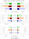
The idea of a tissue engineered nucleus implant is to seed cells in a three-dimensional collagen matrix. This matrix may serve as a scaffold for a tissue engineered nucleus implant. The aim of this study was to investigate whether implantation of the collagen matrix into a spinal segment after nucleotomy is able to restore disc height and flexibility. The implant basically consists of condensed collagen type-I matrix. For clinical use, this matrix will be used for reinforcing and supporting the culturing of nucleus cells. In experiments, matrixes were concentrated with barium sulfate for X-ray purposes and cell seeding was disclaimed in order to evaluate the biomechanical performance of the collagen material. Six bovine lumbar functional spinal units, aging between 5 and 6 months, were used for the biomechanical in-vitro test. In each specimen, an oblique incision was performed, the nucleus was removed and replaced by a collagen-type-I matrix. Specimens were mounted in a custom-built spine tester, and subsequently exposed to pure moments of 7.5 Nm to move within the three anatomical planes. Each tested stage (intact, nucleotomy and implanted) was evaluated for range of motion, neutral zone and change in disc height. Removal of the nucleus significantly reduced disc height by 0.84 mm in respect to the intact stage and caused an instability in the segment. Through the implantation of the tissue engineered nucleus it was possible to restore this height and stability loss, and even to increase slightly the disc height of 0.07 mm compared with the intact stage. There was no statistical difference between the stability provided by the implant and intact stage. Results of movements in lateral bending and axial rotation showed the same trend compared to flexion/extension. However, implant extrusions have been observed in three of six cases during the flexibility assessment. The results of this study directly reflect the efficacy of vital nucleus replacement to restore disc height and to provide stability to intervertebral discs. However, from a biomechanical point of view, the challenge is to employ an appropriate annulus fibrosus sealing method, which is capable to keep the nucleus implant in place over a long-time period. Securing the nucleus implant inside the disc is one of the most important biomechanical prerequisites if such a tissue engineered implant shall have a chance for clinical application.
Figures


Similar articles
-
Nucleus replacement could get a new chance with annulus closure.Eur Spine J. 2020 Jul;29(7):1733-1741. doi: 10.1007/s00586-020-06419-2. Epub 2020 Apr 24. Eur Spine J. 2020. PMID: 32333186
-
Total disc replacement using a tissue-engineered intervertebral disc in vivo: new animal model and initial results.Evid Based Spine Care J. 2010 Aug;1(2):62-6. doi: 10.1055/s-0028-1100918. Evid Based Spine Care J. 2010. PMID: 23637671 Free PMC article.
-
Regeneration of the intervertebral disc with nucleus pulposus cell-seeded collagen II/hyaluronan/chondroitin-6-sulfate tri-copolymer constructs in a rabbit disc degeneration model.Spine (Phila Pa 1976). 2011 Dec 15;36(26):2252-9. doi: 10.1097/BRS.0b013e318209fd85. Spine (Phila Pa 1976). 2011. PMID: 21358466
-
Injectable biomaterials and vertebral endplate treatment for repair and regeneration of the intervertebral disc.Eur Spine J. 2006 Aug;15 Suppl 3(Suppl 3):S414-21. doi: 10.1007/s00586-006-0172-2. Epub 2006 Jul 26. Eur Spine J. 2006. PMID: 16868785 Free PMC article. Review.
-
Cell transplantation in lumbar spine disc degeneration disease.Eur Spine J. 2008 Dec;17 Suppl 4(Suppl 4):492-503. doi: 10.1007/s00586-008-0750-6. Epub 2008 Nov 13. Eur Spine J. 2008. PMID: 19005697 Free PMC article. Review.
Cited by
-
In vivo biofunctional evaluation of hydrogels for disc regeneration.Eur Spine J. 2014 Jan;23(1):19-26. doi: 10.1007/s00586-013-2998-8. Eur Spine J. 2014. PMID: 24121748 Free PMC article.
-
Percutaneous posterolateral approach for the simulation of a far-lateral disc herniation in an ovine model.Eur Spine J. 2018 Jan;27(1):222-230. doi: 10.1007/s00586-017-5362-6. Epub 2017 Oct 27. Eur Spine J. 2018. PMID: 29080003
-
Multiscale Regulation of the Intervertebral Disc: Achievements in Experimental, In Silico, and Regenerative Research.Int J Mol Sci. 2021 Jan 12;22(2):703. doi: 10.3390/ijms22020703. Int J Mol Sci. 2021. PMID: 33445782 Free PMC article. Review.
-
Fabrication, maturation, and implantation of composite tissue-engineered total discs formed from native and mesenchymal stem cell combinations.Acta Biomater. 2020 Sep 15;114:53-62. doi: 10.1016/j.actbio.2020.05.039. Epub 2020 Jun 4. Acta Biomater. 2020. PMID: 32505801 Free PMC article.
-
[Biomechanics of the intervertebral disc : Consequences of degenerative changes].Orthopadie (Heidelb). 2024 Dec;53(12):912-917. doi: 10.1007/s00132-024-04578-4. Epub 2024 Nov 5. Orthopadie (Heidelb). 2024. PMID: 39499289 Review. German.
References
-
- Alini M, Li W, Markovic P, Aebi M, Spiro RC, and Roughley PJ (2003) The potential and limitations of a cell-seeded collagen/hyaluronan scaffold to engineer an intervertebral disc-like matrix. Spine 28(5):446–454; discussion 453 - PubMed
MeSH terms
Substances
LinkOut - more resources
Full Text Sources
Other Literature Sources
Medical

