. 2006 Oct;17(10):4300-17.
doi: 10.1091/mbc.e06-05-0421. Epub 2006 Jul 26.

A single common portal for clathrin-mediated endocytosis of distinct cargo governed by cargo-selective adaptors

Affiliations

A single common portal for clathrin-mediated endocytosis of distinct cargo governed by cargo-selective adaptors

Peter A Keyel et al. Mol Biol Cell. 2006 Oct.

Abstract

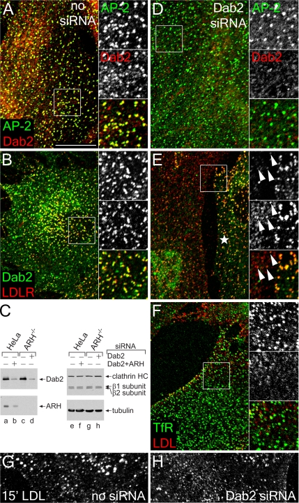
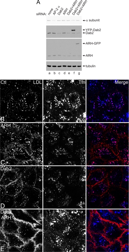
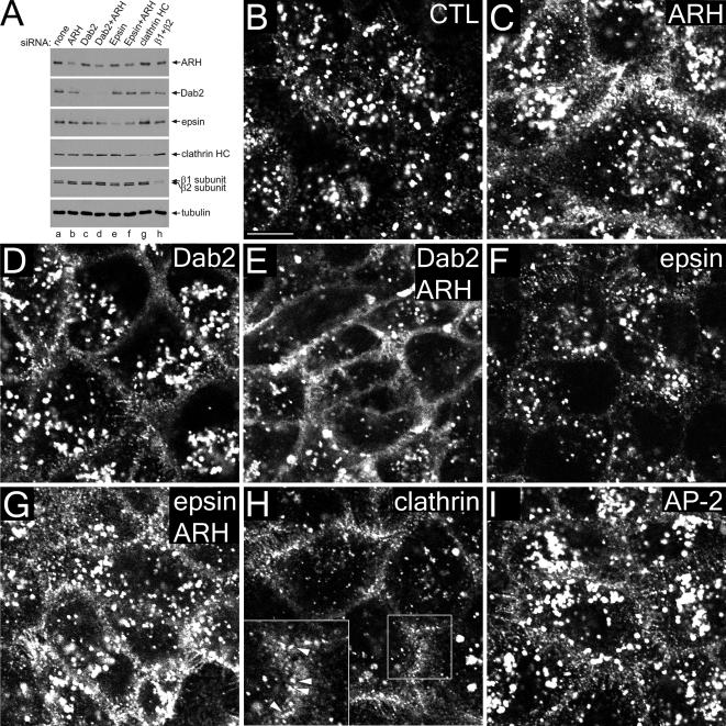
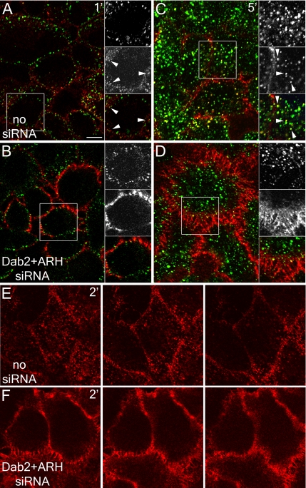
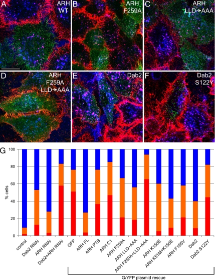
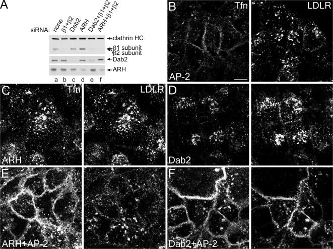
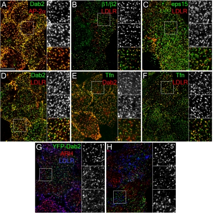
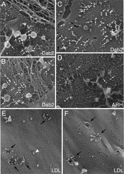
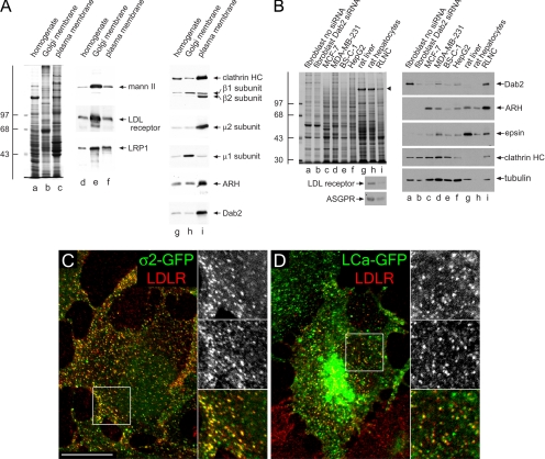
Sorting of transmembrane cargo into clathrin-coated vesicles requires endocytic adaptors, yet RNA interference (RNAi)-mediated gene silencing of the AP-2 adaptor complex only disrupts internalization of a subset of clathrin-dependent cargo. This suggests alternate clathrin-associated sorting proteins participate in cargo capture at the cell surface, and a provocative recent proposal is that discrete endocytic cargo are sorted into compositionally and functionally distinct clathrin coats. We show here that the FXNPXY-type internalization signal within cytosolic domain of the LDL receptor is recognized redundantly by two phosphotyrosine-binding domain proteins, Dab2 and ARH; diminishing both proteins by RNAi leads to conspicuous LDL receptor accumulation at the cell surface. AP-2-dependent uptake of transferrin ensues relatively normally in the absence of Dab2 and ARH, clearly revealing delegation of sorting operations at the bud site. AP-2, Dab2, ARH, transferrin, and LDL receptors are all present within the vast majority of clathrin structures at the surface, challenging the general existence of specialized clathrin coats for segregated internalization of constitutively internalized cargo. However, Dab2 expression is exceptionally low in hepatocytes, likely accounting for the pathological hypercholesterolemia that accompanies ARH loss.


Figures

Figure 1.
Figure 1.
Dab2 activity in ARH-null fibroblasts. GM00697 ARH−/− fibroblasts nucleofected with mock (A, B, and G) or Dab2-specific siRNA duplexes (D–F and H) were fixed and stained with anti-AP-2 α subunit mAb AP.6 and/or affinity-purified anti-Dab2 antibodies, or, before fixation, were incubated on ice with anti-LDL receptor mAb IgG-C7 (LDLR), or anti-transferrin receptor (TfR) mAb RVS-10 and DiI-LDL on ice for 60 min. Representative single confocal optical sections are shown with color-separated AP-2 (green channel, top) and Dab2 (red channel, middle) and merged magnifications of each boxed region shown on the right. Alternatively, whole cell lysates from HeLa SS6 or ARH−/− fibroblast transfected with mock, Dab2+ARH, or Dab2 siRNA duplexes were prepared, resolved by SDS-PAGE, and transferred to nitrocellulose (C). Portions of the blots were probed with anti-Dab2 or ARH polyclonal antibodies, anti-clathrin heavy chain (HC) mAb TD.1 and anti-β1/β2 subunit mAb 100/1, or anti-tubulin mAb E7, and only the relevant region of each blot is shown. Mock (G) or Dab2 siRNA transfected (H) ARH−/− fibroblast cells were also incubated with DiI-LDL at 37°C for 15 min before fixation. In E, an untransfected cell is indicated with an asterisk, and residual Dab2-positive structures containing LDL receptor are shown with arrowheads. Scale bar, 10 μm.
Figure 2.
Figure 2.
Cargo selective effect of Dab2 and ARH gene silencing. HeLa SS6 cells transiently transfected with mock (A, lane a, and B) or siRNA duplexes targeting the AP-2 α subunit (A, lane b), Dab2 (A, lane c, and D), ARH (A, lane d, and C), or Dab2+ARH alone (A, lane e, and E), or together with YFP-Dab2 (A, lane f) or ARH-GFP (A, lane g) were lysed, resolved by SDS-PAGE, and transferred to nitrocellulose. Portions of the blots were probed with anti-AP-2 α subunit mAb clone 8, anti-Dab2 or anti-ARH polyclonal antibodies, or anti-tubulin mAb E7 (A). Only the relevant portions of the blots are shown. Alternatively, transfected cells on coverslips were incubated together with 4 μg/ml DiI-LDL (left, red in merge) and 50 μg/ml Tfn633 (center, blue in merge) for 15 min at 37°C and fixed (B–E). Representative single confocal optical sections are shown. Note the pronounced surface accumulation of LDL, but not transferrin, in the Dab2+ARH knockdown and to a much lesser extent in the single knockdowns. Scale bar, 10 μm.
Figure 3.
Figure 3.
Dab2 and ARH are LDL-specific CLASPs. HeLa SS6 cells transiently transfected with mock (A, lane a, and B) or with specific siRNA duplexes directed against ARH (A, lane b, and C), Dab2 (A, lane c, and D), Dab2+ARH (A, lane d, and E), epsin 1 (A, lane e, and F), epsin1+ARH (A, lane f, and G), clathrin heavy chain (A, lane g, and H), or AP-1 β1 and AP-2 β2 subunits (A, lane h, and I) were lysed, resolved by SDS-PAGE, and transferred to nitrocellulose. Portions of the blots were probed with either affinity-purified anti-ARH, anti-Dab2, or anti-epsin 1, anti-β1/β2 subunit mAb 100/1 antibodies, anti-clathrin HC mAb TD.1, or anti-tubulin mAb E7. Alternatively, transfected cells on coverslips were incubated with 4 μg/ml DiI-LDL for 15 min at 37°C and fixed (B–I). Representative single confocal optical sections are shown with a magnification of the boxed region in H shown on the lower left. Note the dramatic surface accumulation and diminished internalization of LDL in clathrin or Dab2+ARH double knockdowns, but some surface LDL still appears clustered in puncta in the absence of clathrin (H inset, arrowheads). Scale bar, 10 μm.
Figure 4.
Figure 4.
Slowed LDL uptake in Dab2- and ARH-depleted HeLa cells. (A–D) HeLa SS6 cells either mock transfected (A and C) or transfected with Dab2+ARH siRNA duplexes (B and D) were incubated at 37°C in the continuous presence of both 25 μg/ml Tfn488 (green) and 5 μg/ml DiI-LDL (red) either for 1 (A and B) or 5 min (C and D), rapidly washed on ice, and then fixed. The boxed regions are enlarged at the right of each image, with transferrin (green), LDL (red) and merged channels shown. Arrowheads mark punctate endosomal structures. Importantly, the DiI-LDL concentration used is considerably lower than the ∼50 μg/ml required to saturate the surface LDL receptor in control cells (Brown and Goldstein, 1986) and, consequently, not all LDL receptors are occupied under these conditions. (E and F) HeLa SS6 cells either mock transfected (E) or transfected with Dab2+ARH siRNA duplexes (F) were incubated at 37°C with 5 μg/ml DiI-LDL for 2 min, washed on ice, and fixed. Representative sequential confocal optical sections are shown to illustrate the general delay in LDL internalization in the Dab2+ARH-depleted cells. Scale bar, 10 μm.
Figure 5.
Figure 5.
Cargo- and AP-2–binding domains of ARH and Dab2 are necessary for LDL sorting. HeLa SS6 cells transiently transfected with siRNAs targeting both ARH and Dab2 along with either wild-type ARH-GFP (A), or F259A (B), 212LLD→AAA (C), F259A+212LLD→AAA (D) ARH mutations, or wild-type YFP-Dab2 (E) or a S122Y Dab2 mutation (F), were grown in DMEM with LPDS overnight, incubated with 4 μg/ml DiI-LDL (red) and 50 μg/ml Tfn633 (blue) for 15 min at 37°C, and then fixed. Scale bar, 10 μm. (G) Quantitation of various ARH-GFP or YFP-Dab2 constructs rescuing LDL internalization. The percentage of cells showing normal (blue), impaired (approximating the level of LDL internalization seen in ARH or Dab2 single knockdowns; orange), or blocked LDL internalization (red) was determined for control (n = 1194 cells), Dab2 (n = 718), ARH (n = 686) or Dab2+ARH (n = 3455) siRNA-treated cells or for the Dab2+ARH siRNA background with rescue plasmids GFP (n = 412), ARH full-length (ARH FL, n = 231), ARH residues 1–179 (ARH PTB, n = 195), ARH residues 180–308 (ARHC1, n = 177), ARH F259A (n = 235), ARH LLD→AAA (n = 93), ARH F259A+LLD→AAA (n = 178), ARH K150E (n = 20), ARH K51M+K150M (n = 91), ARH F156V (n = 110), Dab2 (n = 183), or Dab2 S122Y (n = 45).
Figure 6.
Figure 6.
ARH requires AP-2 for LDL-sorting function. HeLa SS6 cells mock-transfected (A, lane a) or transfected with AP-2 (β1+β2 subunits; A, lane b, and B), Dab2 (A, lane c, and D), ARH (A, lane d, and C), or Dab2+β1+β2 (A, lane e, and F) or ARH+β1+β2 (A, lane f, and E) specific siRNA duplexes grown overnight in DMEM with 10% LPDS were lysed, resolved by SDS-PAGE and transferred to nitrocellulose. Portions of replicate blots were probed with anti-clathrin heavy-chain mAb TD.1, anti-β1/β2 subunit mAb 100/1, or polyclonal anti-Dab2 or anti-ARH antibodies (A). Alternatively, transfected cells on coverslips were incubated for 15 min at 37°C with 25 μg/ml Tfn568 (Tfn) and anti-LDL receptor mAb IgG-C7 (LDLR) before fixing (B–F). Representative single confocal optical sections are shown. Scale bar, 10 μm.
Figure 7.
Figure 7.
General localization of CLASPs and LDL receptors to endocytic coated coats at steady state. HeLa SS6 cells cultured in DMEM containing 10% LPDS overnight were either fixed and stained with anti-Dab2 antibody and anti-AP-2 α subunit mAb AP.6 (A), or surface-labeled with anti-LDL receptor mAb IgG-C7 (LDLR), fixed and stained with either affinity-purified anti-AP-1/2 β1/β2-subunit GD/1 (B), anti-eps15 (C), or anti-Dab2 (D) antibodies, or surface-labeled with Tfn488 (Tfn), fixed and stained with anti-Dab2 antibody (E), or surface-labeled with both Tfn488 and anti-LDL receptor mAb IgG-C7 (F). Representative single confocal optical sections are shown with color-separated and merged magnifications of each boxed region shown on the right. Alternatively, HeLa SS6 cells transfected with YFP-Dab2 were incubated with anti-LDL receptor mAb IgG-C7 for 1 (G) or 5 min (H) at 37°C, fixed, and stained with anti-ARH antibody. Scale bar, 10 μm.
Figure 8.
Figure 8.
Ultrastructural localization of Dab2, ARH, and LDL in clathrin coats. (A–F) Fixed plasma membrane sheets prepared from control GM01386 fibroblasts (A, D, and E) or HeLa cells (B, C, and F) were labeled with either affinity-purified anti-Dab2 or ARH antibodies, and then with secondary antibodies conjugated to 15-nm colloidal-gold particles. Individual gold particles appear as white spheres (arrows), and representative freeze-etch images show the spatial distribution of endogenous Dab2 and ARH. (E and F) Control fibroblasts (GM01386, E) or HeLa cells (F) incubated in 10% LPDS for 24 h were incubated with 50 μg/ml LDL at either 0°C (F) or 37°C (E) and fixed before preparation of replicas of the external cell surface. Individual LDL particles (arrowheads) and invaginated pits (arrows) are indicated.
Figure 9.
Figure 9.
Live cell dynamics of Dab2 and ARH. HeLa cells transiently transfected with either YFP-Dab2 (A) or ARH-GFP (B) were photobleached (boxed region) with a 405-nm laser at time 0 and imaged every 5.2 s to follow recovery of the fluorescent signal. Magnified views of the bleached region at the indicated times are shown in the insets. (C) Normalized fluorescence intensity of YFP-Dab2 (14 separate cells, ■) and ARH-GFP (29 separate cells, □) within the bleached regions. (D–F) YFP-Dab2 (middle panels, green) transfected HeLa cells were grown in LPDS overnight, incubated with 4 μg/ml DiI-LDL (top panels, red) and imaged every 3.3 (D) or 3.4 s (E and F) either during LDL addition (D) or 21 min later (E and F). (D) Quantitation of the fluorescence intensity of 17 structures during early time points shows that the intensity of LDL (▴) accumulates in these Dab2-positive structures (■). The averaged intensity for Dab2 and LDL in all these structures is shown. (E and F) At later time points, the internalized DiI-LDL has moved to larger, brighter YFP-Dab2-negative endosomal compartments, though LDL still accumulates and internalizes from surface Dab2-positive structures. Importantly, the concentration of LDL used in these experiments is ∼10-fold below the saturating concentration at 37°C (>50 μg/ml; Brown and Goldstein, 1986), so only a subset of surface LDL receptors are marked with fluorescent ligand. A cluster of selected surface YFP-Dab2 structures containing LDL, indicated with the rectangle in E, is magnified in F. Separation of the DiI-LDL signal from the Dab2-YFP and local movement of the internalized vesicle are evident. E and F are from Supplementary Movie 2. The brackets indicate the vertical position from each image set used to generate the kymograph. Scale bar, 10 μm in A and B, 1 μm in E, and 500 nm in F. Kymograph scale bar, 20 s. Note the striking differences in the dynamics of the Dab2-YFP and DiI-LDL structures seen in the kymographs.
Figure 10.
Figure 10.
Cell-type-specific expression of Dab2 in the liver. (A) Samples of 25 μg of rat liver homogenate (lanes a, d, and g), purified rat liver Golgi membranes (lanes b, e, and h), or rat liver plasma membrane sheets (lane c, f and i) were resolved by SDS-PAGE and either stained with Coomassie blue (lane a–c) or transferred to nitrocellulose (lanes d–i). Portions of replicate blots were probed with antibodies directed against α-mannosidase II (mann II), the LDL receptor, LRP1, clathrin heavy chain (HC), AP-1/2 β1/β2 subunits, AP-2 μ2 subunit, AP-1 μ1 subunit, ARH, or Dab2. The position of the molecular mass standards (in kDa) is indicated on the left. (B) Lysates from ARH−/− fibroblasts either mock (lane a) transfected or transfected with Dab2 siRNA (lane b), MCF-7 (lane c), MDA-MB-231 (lane d), BS-C-1 (lane e), or HepG2 (lane f) cultured cell lines, rat liver homogenate (lane g), rat hepatocyte (lane h), or rat liver nonhepatocyte cell (RLNC, lane i) lysates were resolved by SDS-PAGE under reducing (top left, right panels) or nonreducing (bottom left panels) conditions and stained with Coomassie blue (top left) or transferred to nitrocellulose. A major 180-kDa hepatic polypeptide that prevents efficient transfer of the clathrin heavy chain onto nitrocellulose is indicated on the stained gel (arrowhead). Relevant portions of the blots were probed with affinity-purified anti-Dab2, anti-ARH, anti-epsin or rat-specific anti-LDL receptor polyclonal antibodies, or anti-clathrin HC mAb TD.1, anti-tubulin mAb E7, or anti-asialoglycoprotein receptor mAb 8D7. Analogous results are obtained using an independent goat anti-Dab2 antibody (unpublished data). (C and D) BS-C-1 cells stably expressing either AP-2 ς2-GFP (C, green) or clathrin LCa-GFP (D, green) were grown overnight in DMEM containing LPDS, surface labeled with anti-LDL receptor mAb C7 (LDLR, red) on ice. Representative single confocal optical sections of fixed cells are shown with color-separated and merged magnifications of each boxed region shown on the right. Scale bar, 10 μm.

Similar articles

Cited by

References

    1. Ahle S., Mann A., Eichelsbacher U., Ungewickell U. Structural relationships between clathrin assembly proteins from the Golgi and the plasma membrane. EMBO J. 1988;7:919–929. - PMC - PubMed
    1. Ajioka R. S., Kaplan J. Characterization of endocytic compartments using the horseradish peroxidase-diaminobenzidine density shift technique. J. Cell Biol. 1987;104:77–85. - PMC - PubMed
    1. Anderson R. G., Vasile E., Mello R. J., Brown M. S. Immunocytochemical visualization of coated pits and vesicles in human fibroblasts: relation to low density lipoprotein receptor distribution. Cell. 1978;15:919–933. - PubMed
    1. Arca M. Autosomal recessive hypercholesterolaemia in Sardinia, Italy, and mutations in ARH: a clinical and molecular genetic analysis. Lancet. 2002;359:841–847. - PubMed
    1. Barriere H., Nemes C., Lechardeur D., Khan-Mohammad M., Fruh K. Molecular basis of Ub-dependent internalization of membrane proteins in mammalian cells. Traffic. 2006;7:282–297. - PubMed

Publication types

MeSH terms

LinkOut - more resources