. 2006 Jul 26;26(30):7839-48.
doi: 10.1523/JNEUROSCI.1624-06.2006.

Neural stem cells rescue nervous purkinje neurons by restoring molecular homeostasis of tissue plasminogen activator and downstream targets

Affiliations

Neural stem cells rescue nervous purkinje neurons by restoring molecular homeostasis of tissue plasminogen activator and downstream targets

Jianxue Li et al. J Neurosci. .

Abstract

Neural stem cells (NSCs) offer special therapeutic prospects because they can be isolated from the CNS, expanded ex vivo, and re-implanted into diseased CNS where they not only migrate and differentiate according to cues from host tissue but also appear to be capable of affecting host cells. In nervous (nr) mutant mice Purkinje neuron (PN) mitochondria become abnormal by the second postnatal week, and a majority of PNs die in the fourth to fifth weeks. We previously identified in nr cerebellum a 10-fold increase in tissue plasminogen activator (tPA) as a key component of the mechanism causing nr PN death. Here we report that undifferentiated wild-type murine NSCs, when transplanted into the newborn nr cerebellar cortex, do not replace host PNs but contact imperiled PNs and support their mitochondrial function, dendritic growth, and synaptogenesis, subsequently leading to the rescue of host PNs and restoration of motor coordination. This protection of nr PNs also is verified by an in vitro organotypic slice model in which nr cerebellar slices are cocultured with NSCs. Most importantly, the integrated NSCs in young nr cerebellum rectify excessive tPA mRNA and protein to close to normal levels and protect the mitochondrial voltage-dependent anion channel and neurotrophins, downstream targets of the tPA/plasmin proteolytic system. This report demonstrates for the first time that NSCs can rescue imperiled host neurons by rectifying their gene expression, elevating somatic stem cell therapeutic potential beyond solely cell replacement strategy.


Figures

Figure 1.
Figure 1.
NSCs rescued nr cerebellar PNs. A, Neutral red-stained section of young (P14) nr mouse cerebellum displays a normal cortical cytoarchitecture, including the external GL, the ML, the PN layer (PL, between two blue dotted lines), and the internal GL. The right panel is an enlargement of the small green frame in the left panel. B, Cytoarchitecture and neuronal marker expression of adult (P60) nr mouse cerebellum. Cerebellar sections were immunostained with anti-parvalbumin (parv; red) antibody for interneurons in the ML (left), with anti-NeuN (neun; red) antibody for granule cells in the internal GL (right), and with DAPI (dapi; blue) for nuclei. C, PNs in cerebellar vermis and hemispheres of adult (P60) mice. Ca, Cerebellar sections were stained with anti-calbindin (calb; red) antibody for PNs. Representative images show that the PN number obviously was reduced in the cerebellar hemispheres of nr mice and was preserved significantly by NSC transplantation (nr+nsc). Cb, PN numbers in cerebellar vermis and hemispheres of three groups of mice (wt, nr, and nr+nsc). Values represent the means ± SD of PN number (percentage of wt; n = 9; nr vs nr+nsc in hemispheres, p < 0.01). Cc, Western blot shows that the decreased calb in adult nr mouse cerebellum mostly was recovered in nr+nsc cerebellum. D, Host origin of surviving PNs. Da, Cerebellar section of NSC-transplanted nr mouse (P40) was coimmunostained with anti-calb antibody for PNs and anti-β-gal (green) antibody for NSCs. No colocalization of calb and β-gal was found in PNs. Db, Cerebellar section of male NSC-transplanted female nr mice (P40) was stained with DAPI. Barr bodies (yellow arrows) were observed in nuclei of most surviving PNs. E, Relationship between NSC integration and PN survival. Ea, The number of surviving PNs in adult nr+nsc mice (P40–P60) was correlated positively with the number of NSCs integrated in cerebellar parenchyma (red oblique line; r = 0.76; n = 15; p < 0.01), but not with the number of NSCs in the superficial meninges (green oblique line; r = 0.23; n = 15; p > 0.05). Eb, Representative section of adult (P40) nr+nsc mouse cerebellum was stained with X-gal (blue dots). NSCs were counted from three sections of each mouse. Scale bars: A–C, E, 50 μm; Da, 15 μm; Db, 5 μm.
Figure 2.
Figure 2.
Surviving PNs retained their motor function. A, Results of the rotarod test showed that NSC transplantation did not affect motor function of adult wt mice (wt vs wt+nsc) but significantly restored the motor function of adult nr mice (nr vs nr+nsc). Values represent the means ± SD of time spent on the rod (n = 6–8 for each group of three independent tests; ∗∗p < 0.01). B, The improved motor behavior was correlated positively to the number of surviving PNs in NSC-transplanted nr mouse cerebellum (r = 0.83; n = 8 paired samples; p < 0.01).
Figure 3.
Figure 3.
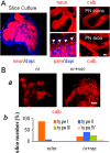
NSCs rescued PNs in nr cerebellar slice cultures. A, Cerebellar slice cultures retained the normal cytoarchitecture of three cortical layers and white matter, i.e., parvalbumin (parv) for the ML, calbindin (calb) for PNs, and NeuN (neun) for the GL. Arrowheads point to basket neurons. B, NSCs rescued PNs in cerebellar slice cultures. Ba, P9 mouse cerebellar slices were cultured for 17 d (P9 + 17 DIV) and stained with anti-calb antibody for PNs. Representative images show that NSCs, when cocultured with cerebellar slices, increased nr PN survival. Bb, Compositions of four types of slices (Marin-Teva et al., 2004) (also see Results) were different between nr cultures and nr+nsc cocultures. For comparison between two kinds of cultures on three independent experiments, p < 0.01. Scale bars: A, B, 50 μm.
Figure 4.
Figure 4.
NSCs approached and contacted host PNs. A, NSCs (bgal; green) in vivo, which had been transplanted into the external GL of newborn nr mouse cerebellum, approached (left) and contacted (right) host PNs (calb; red). B, NSCs (bgal; green) in vitro, which had been cocultured with cerebellar slices of nr mice, migrated into the slices and extensively contacted PNs (calb; red). The two right panels are enlargements of the small yellow frame in the left panel. X represents the section images from the x-axis point of view, and Y represents the section images from the y-axis point of view. Both X and Y images show the points of contact between PN (red) and NSC (green). Scale bars: Aa, 30 μm; Ab, 10 μm; B, 25 μm.
Figure 5.
Figure 5.
NSCs protected nr PN mitochondria, dendrites, and axons. A, PNs in young (P16) mouse cerebellar sections (1 μm thick) were stained with alkaline toluidine blue. Numerous round mitochondria (dark puncta) were found in nr PN soma (red dotted ellipse), but not in wt or nr+nsc PN soma (red dotted ellipse). B, A significant reduction was shown in young (P16) nr PN small dendrites (calb; red) but recovered in nr+nsc PN dendrites in the ML. C, The nr+nsc PNs extended long axons (calb; red) through white matter toward the cerebellar nuclei, but they remained compromised in adult (P60) nr cerebellum that held residual PNs. Scale bars: A, 5 μm; B, 30 μm; C, 50 μm.
Figure 6.
Figure 6.
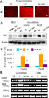
NSCs rectified excessive tPA in nr cerebellum. A, tPA protein levels. Aa, Representative images of young (P16) mouse cerebellum stained with anti-tPA (red) antibody. The tPA level in nr cerebellum was obviously higher than in wt and nr+nsc cerebella. Scale bar, 30 μm. Ab, Levels of tPA protein measured with Western blot were extremely elevated in young (P16) and adult (P120) nr mouse cerebella as compared with those in wt or nr+nsc cerebella. Cultured NSCs in vitro produced very small amounts of tPA protein. Ac, Quantification of tPA protein in young (P16) and adult (P120–P180) cerebella of wt, nr, and nr+nsc mice. Values represent the means ± SD of tPA relative levels (percentage of wt; n = 5). Comparison between nr and nr+nsc, ∗∗p < 0.01. B, tPA(c) and tPA(n) mRNAs were detected by using RT-PCR. Expression of tPA mRNA was upregulated greatly in young (P16) and adult (P120) nr mouse cerebellum and testis, as compared with wt controls. NSC transplantation (nr+nsc) into cerebellum suppressed the increased tPA mRNA in nr cerebellum, but not in nr testis.
Figure 7.
Figure 7.
NSCs diminished the effects of excessive tPA on downstream targets. A, Neurotrophins and postsynaptic marker. Aa, Representative images of young (P14) mouse cerebellar sections costained with anti-calb (red) and anti-BDNF (green) antibodies. BDNF, a downstream target of the tPA/plasmin system, existed in the external GL (in rectangle) of wt and nr+nsc cerebella but was reduced greatly in nr cerebellum. Ab, Representative images of young (P14) mouse cerebellar sections costained with anti-calb (red) and anti-NT3 (green) antibodies. NT3, a downstream target of the tPA/plasmin system, existed normally in PN somata of wt and nr+nsc cerebella but was reduced greatly in nr cerebellum. Ac, PSD-95 (red) staining, a postsynaptic dendritic marker, was decreased in the ML and increased in PN somata (parallel lines) of young (P16) nr cerebellum as compared with wt and nr+nsc cerebella. Ad, PSD-95 (red) staining showed many fewer postsynaptic connections (arrowheads) between basket cell axons and PN somata (parallel lines) in adult (P60) nr cerebellum as compared with wt and nr+nsc cerebella. B, NSCs in monolayer cultures obviously expressed foxa2 and gbx2 transcription factors. Ba, Representative images of NSC cultures in undifferentiated phase (undif. nsc) and differentiated phase (dif. nsc). Bb, Results of RT-PCR with the use of two pairs of primers for each factor showed that foxa2 (primer I for 392 bp and primer II for 349 bp) was expressed equally in undifferentiated and differentiated NSCs, whereas gbx2 products (primer I for 462 bp and primer II for 363 bp) were higher in differentiated NSCs than in undifferentiated NSCs. Scale bars: Aa, 30 μm; Ab, 50 μm; Ac, Ad, 15 μm.

Similar articles

Cited by

References

    1. Banerjee J, Ghosh S (2004). Interaction of mitochondrial voltage-dependent anion channel from rat brain with plasminogen protein leads to partial closure of the channel. Biochim Biophys Acta 1663:6–8. - PubMed
    1. Chen ZL, Strickland S (1997). Neuronal death in the hippocampus is promoted by plasmin-catalyzed degradation of laminin. Cell 91:917–925. - PubMed
    1. Christopherson KS, Ullian EM, Stokes CC, Mullowney CE, Hell JW, Agah A, Lawler J, Mosher DF, Bornstein P, Barres BA (2005). Thrombospondins are astrocyte-secreted proteins that promote CNS synaptogenesis. Cell 120:421–433. - PubMed
    1. Crompton M (1999). The mitochondrial permeability transition pore and its role in cell death. Biochem J 341:233–249. - PMC - PubMed
    1. De Jager PL, Harvey D, Polydorides AD, Zuo J, Heintz N (1998). A high-resolution genetic map of the nervous locus on mouse chromosome 8. Genomics 48:346–353. - PubMed

Publication types

Substances

LinkOut - more resources