

Stress-induced alterations in prefrontal cortical dendritic morphology predict selective impairments in perceptual attentional set-shifting
- PMID: 16870732
- PMCID: PMC6674229
- DOI: 10.1523/JNEUROSCI.1184-06.2006
Stress-induced alterations in prefrontal cortical dendritic morphology predict selective impairments in perceptual attentional set-shifting
Abstract
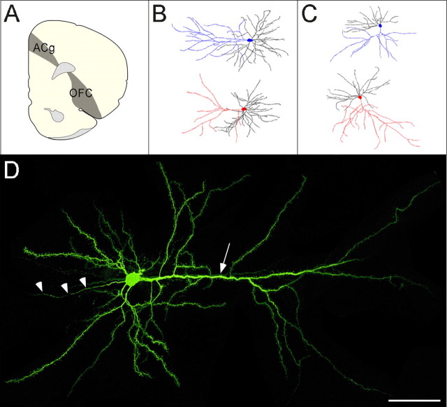
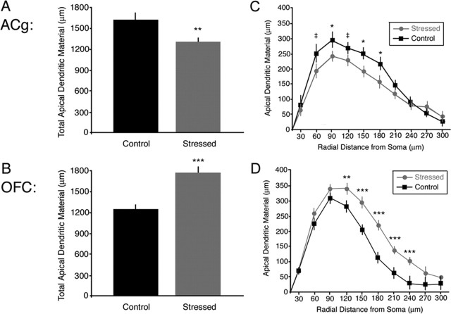
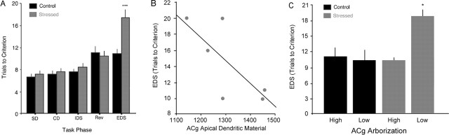
Stressful life events have been implicated clinically in the pathogenesis of mental illness, but the neural substrates that may account for this observation remain poorly understood. Attentional impairments symptomatic of these psychiatric conditions are associated with structural and functional abnormalities in a network of prefrontal cortical structures. Here, we examine whether chronic stress-induced dendritic alterations in the medial prefrontal cortex (mPFC) and orbital frontal cortex (OFC) underlie impairments in the behaviors that they subserve. After 21 d of repeated restraint stress, rats were tested on a perceptual attentional set-shifting task, which yields dissociable measures of reversal learning and attentional set-shifting, functions that are mediated by the OFC and mPFC, respectively. Intracellular iontophoretic injections of Lucifer yellow were performed in a subset of these rats to examine dendritic morphology in layer II/III pyramidal cells of the mPFC and lateral OFC. Chronic stress induced a selective impairment in attentional set-shifting and a corresponding retraction (20%) of apical dendritic arbors in the mPFC. In stressed rats, but not in controls, decreased dendritic arborization in the mPFC predicted impaired attentional set-shifting performance. In contrast, stress was not found to adversely affect reversal learning or dendritic morphology in the lateral OFC. Instead, apical dendritic arborization in the OFC was increased by 43%. This study provides the first direct evidence that dendritic remodeling in the prefrontal cortex may underlie the functional deficits in attentional control that are symptomatic of stress-related mental illnesses.
Figures
Similar articles
-
Structural and functional alterations to rat medial prefrontal cortex following chronic restraint stress and recovery.Neuroscience. 2009 Dec 1;164(2):798-808. doi: 10.1016/j.neuroscience.2009.08.053. Epub 2009 Aug 29. Neuroscience. 2009. PMID: 19723561 Free PMC article.
-
Reversibility of apical dendritic retraction in the rat medial prefrontal cortex following repeated stress.Exp Neurol. 2005 Nov;196(1):199-203. doi: 10.1016/j.expneurol.2005.07.008. Epub 2005 Aug 10. Exp Neurol. 2005. PMID: 16095592
-
NMDA receptor blockade alters stress-induced dendritic remodeling in medial prefrontal cortex.Cereb Cortex. 2011 Oct;21(10):2366-73. doi: 10.1093/cercor/bhr021. Epub 2011 Mar 7. Cereb Cortex. 2011. PMID: 21383235 Free PMC article.
-
Neurochemical modulation of stress-induced cognitive inflexibility in a rat model of an attentional set-shifting task.Pharmacol Rep. 2013;65(6):1479-88. doi: 10.1016/s1734-1140(13)71508-1. Pharmacol Rep. 2013. PMID: 24552995 Review.
-
Stress-induced dendritic remodeling in the medial prefrontal cortex: effects of circuit, hormones and rest.Brain Res. 2009 Oct 13;1293:108-13. doi: 10.1016/j.brainres.2009.03.062. Epub 2009 Apr 8. Brain Res. 2009. PMID: 19361488 Free PMC article. Review.
Cited by
-
Age-dependent effects of repeated amphetamine exposure on working memory in rats.Behav Brain Res. 2013 Apr 1;242:84-94. doi: 10.1016/j.bbr.2012.12.044. Epub 2013 Jan 3. Behav Brain Res. 2013. PMID: 23291159 Free PMC article.
-
The stressed synapse: the impact of stress and glucocorticoids on glutamate transmission.Nat Rev Neurosci. 2011 Nov 30;13(1):22-37. doi: 10.1038/nrn3138. Nat Rev Neurosci. 2011. PMID: 22127301 Free PMC article. Review.
-
Self-affirmation improves problem-solving under stress.PLoS One. 2013 May 1;8(5):e62593. doi: 10.1371/journal.pone.0062593. Print 2013. PLoS One. 2013. PMID: 23658751 Free PMC article.
-
Synaptic Cytoskeletal Plasticity in the Prefrontal Cortex Following Psychostimulant Exposure.Traffic. 2015 Sep;16(9):919-40. doi: 10.1111/tra.12295. Epub 2015 Jun 1. Traffic. 2015. PMID: 25951902 Free PMC article. Review.
-
Guanfacine's mechanism of action in treating prefrontal cortical disorders: Successful translation across species.Neurobiol Learn Mem. 2020 Dec;176:107327. doi: 10.1016/j.nlm.2020.107327. Epub 2020 Oct 17. Neurobiol Learn Mem. 2020. PMID: 33075480 Free PMC article. Review.
References
-
- Casey BJ, Tottenham N, Fossella J (2002). Clinical, imaging, lesion, and genetic approaches toward a model of cognitive control. Dev Psychobiol 40:237–254. - PubMed
-
- Caspi A, Sugden K, Moffitt TE, Taylor A, Craig IW, Harrington H, McClay J, Mill J, Martin J, Braithwaite A, Poulton R (2003). Influence of life stress on depression: moderation by a polymorphism in the 5-HTT gene. Science 301:386–389. - PubMed
-
- Cohen JD, Servan-Schreiber D (1992). Context, cortex and dopamine: a Connectionist approach to behavior and biology in schizophrenia. Psychol Rev 99:47. - PubMed
Publication types
MeSH terms
Grants and funding
LinkOut - more resources
Full Text Sources
Other Literature Sources
Medical