Comparative Study
. 2006 Aug;50(8):2608-20.
doi: 10.1128/AAC.00181-06.

Naturally processed dermcidin-derived peptides do not permeabilize bacterial membranes and kill microorganisms irrespective of their charge

Affiliations
Free PMC article
Comparative Study

Naturally processed dermcidin-derived peptides do not permeabilize bacterial membranes and kill microorganisms irrespective of their charge

H Steffen et al. Antimicrob Agents Chemother. 2006 Aug.
Free PMC article

Abstract

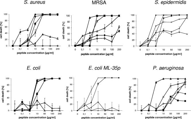
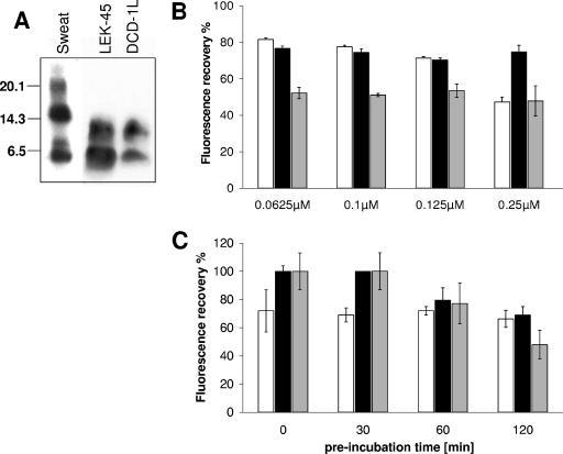
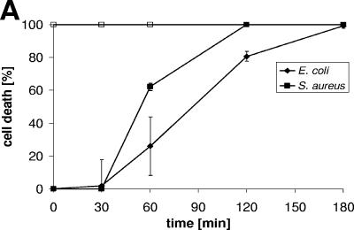
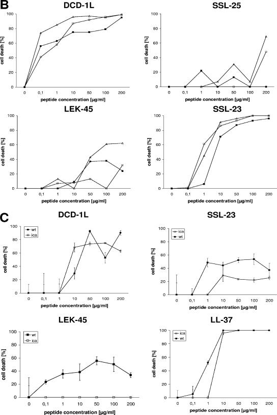
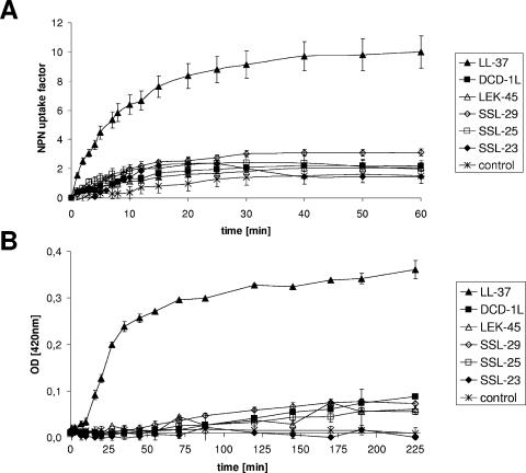
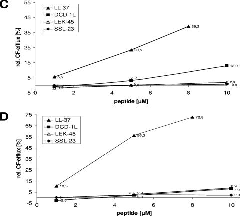
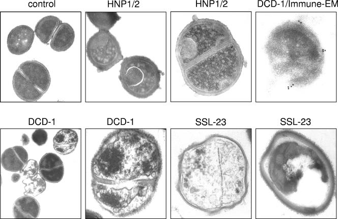
Dermcidin (DCD) is a recently described antimicrobial peptide, which is constitutively expressed in eccrine sweat glands and transported via sweat to the epidermal surface. By postsecretory proteolytic processing in sweat the dermcidin protein gives rise to several truncated DCD peptides which differ in length and net charge. In order to understand the mechanism of antimicrobial activity, we analyzed the spectrum of activity of several naturally processed dermcidin-derived peptides, the secondary structure in different solvents, and the ability of these peptides to interact with or permeabilize the bacterial membrane. Interestingly, although all naturally processed DCD peptides can adopt an alpha-helical conformation in solvents, they have a diverse and partially overlapping spectrum of activity against gram-positive and gram-negative bacteria. This indicates that the net charge and the secondary structure of the peptides are not important for the toxic activity. Furthermore, using carboxyfluorescein-loaded liposomes, membrane permeability studies and electron microscopy we investigated whether DCD peptides are able to permeabilize bacterial membranes. The data convincingly show that irrespective of charge the different DCD peptides are not able to permeabilize bacterial membranes. However, bacterial mutants lacking specific cell envelope modifications exhibited different susceptibilities to killing by DCD peptides than wild-type bacterial strains. Finally, immunoelectron microscopy studies indicated that DCD peptides are able to bind to the bacterial surface; however, signs of membrane perturbation were not observed. These studies indicate that DCD peptides do not exert their activity by permeabilizing bacterial membranes.


Figures

FIG. 1.
FIG. 1.
Antimicrobial activity of DCD-derived peptides against several bacterial strains. The concentration-dependent antimicrobial activity of the DCD-derived peptides DCD-1L (▪), LEK-45 (▵), SSL-29 (⋄), SSL-25 (+), SSL-23 (♦), and LL-37 (▴) on the bacterial strains S. aureus, MRSA, S. epidermidis, E. coli, E. coli ML-35p, and Pseudomonas aeruginosa after 2 to 3 h of incubation in 10 mM phosphate buffer-10 mM NaCl (pH 7.0) is presented. The number of bacterial colonies were counted, and the percentage of cell death calculated as described previously (37). The microbicidal activity was expressed as [1 − (cell survival after peptide incubation)/(cell survival after control peptide incubation)] × 100, which represents the percentage killing of the cells.
FIG. 2.
FIG. 2.
Determination of the oligomerization of the peptides DCD-1L, LEK-45, SSL-23, and LL-37 in solution. (A) Sodium dodecyl sulfate-polyacrylamide gel electrophoresis and Western blot analysis of human eccrine sweat and 4 μg of the DCD peptides LEK-45 and DCD-1L dissolved in water using a polyclonal anti-DCD antibody, which detects the C terminus of DCD-1L. Seen are SDS-stable dimers for LEK-45 and DCD-1L and higher oligomers in sweat. (B) Percent fluorescence recovery of FITC-labeled peptides LL-37 (□), DCD1L (▪), and SSL-23 (░⃞) in 1× PBS (pH 7.4) at different concentrations (0.0625 to 0.25 μM). Peptides were preincubated 2 h in PBS before proteinase K (10 μg/ml) treatment. Oligomerization of the peptides in solution was determined by fluorescence dequenching. (C) Determination of the time-kinetics of oligomerization: peptides (0.25 μM) were incubated for different time points (0 to 120 min) at room temperature in PBS before proteinase K treatment, and the percentage of fluorescence recovery was determined.
FIG.3.
FIG.3.
Time kinetics and antimicrobial activity of DCD-derived peptides against bacterial cell envelope mutants. (A) Time-dependent killing of S. aureus (▪) and E. coli (⧫) by DCD-1L using the CFU assay. Bacteria in the mid-logarithmic phase of growth were incubated with DCD-1L (200 μg/ml, black symbols) at different time intervals (0-180 min). The open squares indicate the antimicrobial activity of the control peptide LL-37 (100 μg/ml). (B) S. aureus cell envelope mutants mprF (□) and dltA (▵) and wild-type SA113 (▪) were incubated with various concentrations of peptides (0.1 to 200 μg/ml) in 10 mM phosphate buffer-10 mM NaCl (pH 7.0) for 2 to 3 h at 37°C. Aliquots of bacterial suspensions were diluted and plated in triplicate on blood agar. The percentage of cell death was determined as described above. (C) S. epidermdis Δica and wild-type S. epidermidis 1457 were incubated with various concentrations of peptides (0.1 to 200 μg/ml) in 10 mM phosphate buffer-10 mM NaCl (pH 7.0) for 2 to 3 h at 37°C, and the percentage of cell death was determined as described above.
FIG.3.
FIG.3.
Time kinetics and antimicrobial activity of DCD-derived peptides against bacterial cell envelope mutants. (A) Time-dependent killing of S. aureus (▪) and E. coli (⧫) by DCD-1L using the CFU assay. Bacteria in the mid-logarithmic phase of growth were incubated with DCD-1L (200 μg/ml, black symbols) at different time intervals (0-180 min). The open squares indicate the antimicrobial activity of the control peptide LL-37 (100 μg/ml). (B) S. aureus cell envelope mutants mprF (□) and dltA (▵) and wild-type SA113 (▪) were incubated with various concentrations of peptides (0.1 to 200 μg/ml) in 10 mM phosphate buffer-10 mM NaCl (pH 7.0) for 2 to 3 h at 37°C. Aliquots of bacterial suspensions were diluted and plated in triplicate on blood agar. The percentage of cell death was determined as described above. (C) S. epidermdis Δica and wild-type S. epidermidis 1457 were incubated with various concentrations of peptides (0.1 to 200 μg/ml) in 10 mM phosphate buffer-10 mM NaCl (pH 7.0) for 2 to 3 h at 37°C, and the percentage of cell death was determined as described above.
FIG.4.
FIG.4.
Effect of DCD-derived peptides on membrane permeability. (A) Outer membrane permeability measured by peptide-mediated NPN uptake in E. coli ML-35p. E. coli cells were incubated with 10 μM NPN in the presence of various concentrations of DCD peptides in 5 mM sodium HEPES buffer (pH 7.4). Enhanced uptake due to membrane permeability was measured by an increase in fluorescence intensity (Ex350 and Em460) caused by partition of NPN into the hydrophobic interior of the outer membrane. At time point 0 min, intact E. coli ML-35p cells were added to the peptides. The results are expressed as NPN uptake factor of fluorescence in arbitrary units. All analyses were performed in triplicates. (B) Inner membrane permeability measured as the influx of ONPG in E. coli ML-35p after the addition of DCD peptides. Stationary-growth-phase E. coli were incubated for 3 h at room temperature in 10 mM NaP (pH 7.0) with 1.67 mM ONPG. The release of ONP by cytoplasmic β-galactosidase was spectrophotometrically monitored at 420 nm. In the reference cuvette, peptides were placed in solvent. All samples were analyzed in triplicates. (C and D) Influence of the peptides DCD-1L (▪), LEK-45 (▵), and SSL-23 (⧫) on CF efflux of unilamellar liposomes made of DOPC (C) and DOPC-DOPG (1:1 molar ratio) (D). Release was determined 4 min after peptide addition at concentrations of 1 to 10 μM. At 4 min the amount of leakage reached a plateau when liposomes still contained a significant amount of CF. Reaction progress was expressed as the percentage of CF released relative to the total fluorescence released after the addition of Triton X-100 solution at the end of each experiment.
FIG.4.
FIG.4.
Effect of DCD-derived peptides on membrane permeability. (A) Outer membrane permeability measured by peptide-mediated NPN uptake in E. coli ML-35p. E. coli cells were incubated with 10 μM NPN in the presence of various concentrations of DCD peptides in 5 mM sodium HEPES buffer (pH 7.4). Enhanced uptake due to membrane permeability was measured by an increase in fluorescence intensity (Ex350 and Em460) caused by partition of NPN into the hydrophobic interior of the outer membrane. At time point 0 min, intact E. coli ML-35p cells were added to the peptides. The results are expressed as NPN uptake factor of fluorescence in arbitrary units. All analyses were performed in triplicates. (B) Inner membrane permeability measured as the influx of ONPG in E. coli ML-35p after the addition of DCD peptides. Stationary-growth-phase E. coli were incubated for 3 h at room temperature in 10 mM NaP (pH 7.0) with 1.67 mM ONPG. The release of ONP by cytoplasmic β-galactosidase was spectrophotometrically monitored at 420 nm. In the reference cuvette, peptides were placed in solvent. All samples were analyzed in triplicates. (C and D) Influence of the peptides DCD-1L (▪), LEK-45 (▵), and SSL-23 (⧫) on CF efflux of unilamellar liposomes made of DOPC (C) and DOPC-DOPG (1:1 molar ratio) (D). Release was determined 4 min after peptide addition at concentrations of 1 to 10 μM. At 4 min the amount of leakage reached a plateau when liposomes still contained a significant amount of CF. Reaction progress was expressed as the percentage of CF released relative to the total fluorescence released after the addition of Triton X-100 solution at the end of each experiment.
FIG. 5.
FIG. 5.
Morphology of peptide-treated S. aureus. Transmission EM and immune-EM of S. aureus (ATCC 25923) treated with either DCD-1, SSL-23, or the alpha-defensins HNP-1 and -2 as a positive control for pore formation in 10 mM sodium phosphate buffer. Bacteria were incubated with 100 μg of DCD-1, SSL-23, and HNP1/2 per ml for 4 h. As a negative control cells were incubated in buffer without peptide. For the immune-EM, bacteria were incubated with a polyclonal antiserum to DCD-1, and the reactivity was detected by immunogold labeling. Seen is the binding of DCD to the bacterial surface.

References

    1. Baechle, D., T. Flad, A. Cansier, H. Steffen, B. Schittek, J. Tolson, T. Herrmann, H. Dihazi, A. Beck, G. A. Mueller, M. Mueller, S. Stevanovic, C. Garbe, C. A. Mueller, and H. Kalbacher. 2006. Cathepsin D is present in human eccrine sweat and involved in the postsecretory processing of DCD-1L. J. Biol. Chem. 281:5406-5415. - PubMed
    1. Beisswenger, C., and R. Bals. 2005. Functions of antimicrobial peptides in host defense and immunity. Curr. Protein Peptide Sci. 6:255-264. - PubMed
    1. Blondelle, S. E., K. Lohner, and M. Aguilar. 1999. Lipid-induced conformation and lipid-binding properties of cytolytic and antimicrobial peptides: determination and biological specificity. Biochim. Biophys. Acta 1462:89-108. - PubMed
    1. Breukink, E., C. van Kraaij, R. A. Demel, R. J. Siezen, O. P. Kuipers, and B. de Kruijff. 1997. The C-terminal region of nisin is responsible for the initial interaction of nisin with the target membrane. Biochemistry 36:6968-6976. - PubMed
    1. Brogden, K. A. 2005. Antimicrobial peptides: pore formers or metabolic inhibitors in bacteria? Nat. Rev. Microbiol. 3:238-250. - PubMed

Publication types

MeSH terms