. 2006 Aug 25;361(4):771-84.
doi: 10.1016/j.jmb.2006.06.048. Epub 2006 Jul 7.

Two distinct binding modes of a protein cofactor with its target RNA

Affiliations

Two distinct binding modes of a protein cofactor with its target RNA

Gregory Bokinsky et al. J Mol Biol. .

Abstract

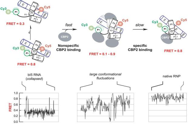
Like most cellular RNA enzymes, the bI5 group I intron requires binding by a protein cofactor to fold correctly. Here, we use single-molecule approaches to monitor the structural dynamics of the bI5 RNA in real time as it assembles with its CBP2 protein cofactor. These experiments show that CBP2 binds to the target RNA in two distinct modes with apparently opposite effects: a "non-specific" mode that forms rapidly and induces large conformational fluctuations in the RNA, and a "specific" mode that forms slowly and stabilizes the native RNA structure. The bI5 RNA folds though multiple pathways toward the native state, typically traversing dynamic intermediate states induced by non-specific binding of CBP2. These results suggest that the protein cofactor-assisted RNA folding involves sequential non-specific and specific protein-RNA interactions. The non-specific interaction potentially increases the local concentration of CBP2 and the number of conformational states accessible to the RNA, which may promote the formation of specific RNA-protein interactions.


Figures

Figure 1
Figure 1
The bI5 construct used for single-molecule FRET experiments and its self splicing reaction. (a) Secondary structure of the bI5 group I intron RNA and its splicing product. Splice sites are marked with purple triangles. The G-U wobble pair and its interaction with the J4/5 junction is marked in red. A biotinylated DNA tether is shown in blue. Splicing was initiated by 2 mM GMP and the resulting close proximity between FRET donor (Cy3) and acceptor (Cy5) in the spliced product leads to a high FRET efficiency, as shown in (b). After adding a stop solution containing EDTA and proteinase K, only the spliced products show FRET = 0.9, while the unspliced RNA molecules show no fluorescence signal due to dissociation of the Cy3-labeled RNA strand in the absence of Mg2+. Inset: Minimal spliced products observed in the absence of GMP.
Figure 2
Figure 2
Similar splicing kinetics for the bI5 RNA as measured by single-molecule and ensemble measurements. (a) Splicing kinetics in 7 mM Mg2+, 50 nM CBP2 and 2 mM GMP determined with single surface-immobilized molecules (open circles, FRET assay, 0.9 ± 0.2 min-1) are the same as those determined for freely diffusing bI5 RNA (closed squares, gel electrophoresis assay, 1.1 ± 0.1 min-1). Nonspecifically bound and excess CBP2 was removed by addition of 100 μg/mL heparin with the 2 mM GMP. (b) Splicing kinetics measured in 40 mM Mg2+, 50 nM CBP2 determined with single surface-immobilized molecules (open circles, 1.0 ± 0.1 min-1) and with freely diffusing bI5 RNA (closed squares, 0.7 ± 0.1 min-1). No heparin was added to remove nonspecifically bound CBP2 in this case. (c) Splicing kinetics for four different bI5 constructs in 40 mM Mg2+, 50 nM CBP2. The splicing rate constant for the Cy3- & Cy5-labeled constructs depicted in Figure 1a (open circles, 1.0 ± 0.1 min-1) is similar to that of a bI5 construct without the Cy3 dye but with Cy5 dye still present on the DNA tether (open squares, 0.7 ± 0.1 min-1) and for a construct omitting the Cy5 dye from the DNA tether (open triangles, 0.6 ± 0.2 min-1). The splicing rate constant of a bI5 construct that lacks the DNA tether is significantly slower (solid triangles, 0.16 ± 0.02 min-1), suggesting that hybridization of the DNA tether to the 3′ exon removes an interfering effect of the 3′ exon on the splicing activity of the intron.
Figure 3
Figure 3
Structural dynamics of the bI5 RNA in the absence of CBP2. (a, b) FRET trajectories of single molecules (left) and a histogram constructed from many bI5 RNA molecules (right) in 7 and 40 mM Mg2+. In constructing the histogram, a threshold is applied to remove FRET values associated with either blinking or photobleaching of Cy5. (c) The dependence of the lower FRET level as a function of Mg2+ concentration for the bI5 (filled triangles) and bI5 ΔP1-P2 (filled squares) constructs. Overlaid is the electrophoretic mobility of the bI5 ΔP1-P2 construct (open circles). (d) Equilibrium constant for the formation of the higher FRET states.
Figure 4
Figure 4
CBP2 binds the bI5 RNA in two modes, each inducing distinct conformational dynamics in the bI5 RNA. (a) Specific binding of CBP2 stabilizes the native structure of the bI5 RNA. Left panel: FRET trajectory of a bI5 RNA specifically bound to CBP2 in 7 mM Mg2+ showing regular transitions between FRET levels of 0.5 and 0.8 (type I binding behavior). Center panel: A FRET histogram of many specific CBP2-bI5 RNA complexes. Right panel: A FRET histogram of U-1C mutant molecules. The regular transitions between FRET = 0.5 and 0.8 are used as a signature for identifying specific binding. (b) Nonspecific binding by CBP2 induces large conformational fluctuations in the bI5 RNA. Left panel: FRET trajectory of a bI5 RNA molecule in 7 mM Mg2+ and 50 nM CBP2, depicting broad and continuous fluctuations across a wide range of FRET values (type II binding behavior). Center panel: FRET histogram obtained from all CBP2-bI5 complexes in 7 mM Mg2+ and 50 nM CBP2. Right panel: Autocorrelation function constructed from many FRET trajectories at 5 laser intensities (I, in arbitrary units).
Figure 5
Figure 5
Concentration dependence of the two CBP2 binding modes. (a) Fractions of RNA molecules not bound to CBP2 (open circles), bound to CBP2 in the type I mode (closed squares) or the type II mode (open triangles) as a function of CBP2 concentration. (b) Fraction of molecules exhibiting the specific (type I) binding mode after incubation with 50 nM CBP2 for 30 min followed by treatment with 100 μg/mL heparin for about 5 min to remove nonspecifically bound CBP2 from the RNA. The data are fit to A[CBP2]n/([CBP2]n + Kn) with a Hill coefficient, n = 1.3 ± 0.3.
Figure 6
Figure 6
CBP2 induces broad structural fluctuations in diverse bI5 RNA variants. (a, b) ΔP1-P2 and ΔP9-P9.1 constructs. The FRET histograms were constructed from many RNA molecules in 7 mM Mg2+ and 50 nM CBP2. (c) The minimum bI5 core with both P1-P2 and P9-P9.1 peripheral domains deleted. A single RNA molecule FRET trajectory in 7 mM Mg2+ and 50 nM CBP2 and A FRET histogram constructed from many molecules are shown. In all cases, the FRET histograms of the constructs in 7 mM Mg2+ in the absence of CBP2 are overlaid for comparison (solid lines).
Figure 7
Figure 7
CBP2 binding destabilize RNA base pairs and causes conformational fluctuations in a RNA hairpin. (a) RNA hairpin construct, SL1, labeled with a fluorescent Cy3 donor and Cy5 acceptor. (Top panels) The FRET histogram of the RNA hairpin indicates that most of the molecules exhibit a well-defined FRET value with a sharp peak at 0.9 as expected for well-formed hairpins. The small peak at FRET ∼ 0.7 may be due to a misfolded form. Upon addition of CBP2, the FRET distribution broadens significantly due to fluctuations toward lower FRET values. A typical trajectory showing such fluctuations is displayed in the lower panel. (b) Control construct, SL1PC, designed to test the potential effect of dye-CBP2 interaction in which the Cy3 and Cy5 dyes are placed on adjacent nucleotides. In this case, CBP2 does not induce broadening of the FRET profile. For clarity, the peak at FRET = 0, reflecting inactive or blinking Cy5 (dashed line), has been subtracted from the experimentally determined histogram. These data were recorded in 10 mM NaCl, 10 mM HEPES pH 7.6, and the oxygen scavenger system as described in Methods.
Figure 8
Figure 8
CBP2 binds rapidly to the bI5 RNA with bimolecular kinetics. (a) Trajectory showing rapid conversion of the free bI5 RNA to CBP2 bound state after addition of 50 nM CBP2 (dotted line). (b) Number of bI5 RNA molecules bound by CBP2 as a function of time. CBP2 binding time is scored as the time when the FRET trajectory shows the broad irregular fluctuations. A single-exponential fit gives a binding rate of 12 min-1. The binding rate constant can also be determined from the average FRET value for an ensemble of bI5 molecules, yielding similar results (data not shown). (c) The binding rate constant as a function of CBP2 concentration.
Figure 9
Figure 9
CBP2-assisted folding of bI5 occurs along multiple pathways that feature dynamic intermediate states. Single-molecule FRET trajectory (a) and histogram (b) illustrating the structural dynamics of a folded bI5 RNA molecule in 40 mM Mg2+ and 50 nM CBP2. A FRET histogram for the U-1C mutant is superimposed as a black line. (c) FRET traces depicting different folding kinetics and intermediate folding states for representative bI5 RNA molecules. The bI5 RNA was pre-incubated at 7 mM Mg2+ prior to buffer-exchange into a folding buffer containing 40 mM Mg2+ and 50 nM CBP2 (dotted line). The upper and lower trajectories were collected at 0.5 Hz time resolution and the middle trajectory was collected at 10 Hz. The molecules often show a dynamic ensemble of intermediates states before attaining the native state, which is identified by a persistent high FRET state dwelling at 0.8 for at least 100 seconds except for brief (1-2 frames) excursions to lower FRET values. Red triangles indicate the attainment of the native state. (d) Fraction of bI5 RNA molecules folded as a function of time after adding the folding buffer containing 40 mM Mg2+ and 50 nM CBP2. The starting conditions are 7 mM Mg2+ (circles) or 40 mM Mg2+ (squares) without CBP2. The folding kinetics at 7 mM Mg2+ were fit to Eq. 1, yielding folding rate constants k1 = 7.2 min-1 and k2 = 0.54 min-1. The overall fraction of molecules folded within the observation time is 65%, among which, the fractions of molecules folded with the faster and slower rates are 47% and 53%, respectively.
Figure 10
Figure 10
Sequential assembly model of the CBP2-bI5 RNA complex. In the absence of protein, the RNA is predominantly in a collapsed state and exhibits transient excursions to a more structured state with native-like features but without catalytic activity. Initial association of CBP2 with the RNA occurs rapidly in a nonspecific mode that increases the local protein concentration and causes substantial conformational fluctuations in the RNA. Subsequent formation of specific CBP2-RNA interactions stabilizes the formation of the native structure of the bI5 RNA. Helices in the bI5 RNA are represented as cylinders. View is looking down the P1 helix. Representative FRET trajectories for each state illustrated in the lower panel are adapted from the data shown in Figures 3a and 9c. Note that the CBP2-bI5 RNP folds along a multitude of pathways with distinct rate constants. In addition, a fraction of molecules misfold and do not attain native states in the observation time window. For clarity, these complex folding kinetics are not displayed in the model.

References

    1. Gesteland RF, Cech TR, Atkins JF. The RNA world. Second Edition Cold Spring Harbor Laboratory Press; New York: 1999.
    1. Pan J, Woodson SA. Folding intermediates of a self-splicing RNA: Mispairing of the catalytic core. J. Mol. Biol. 1998;280:597–609. - PubMed
    1. Pan T, Sosnick TR. Intermediates and kinetics traps in the folding of a large ribozyme revealed by circular dichroism and UV absorbance spectroscopies and catalytic activity. Nat. Struct. Biol. 1997;4:931–938. - PubMed
    1. Russell R, Herschlag D. New pathways in folding of the Tetrahymena group I RNA enzyme. J. Mol. Biol. 1999;291:1155–1167. - PubMed
    1. Treiber DK, Rook MS, Zarrinkar PP, Williamson JR. Kinetic intermediates trapped by native interactions in RNA folding. Science. 1998;279:1943–6. - PubMed

Publication types

LinkOut - more resources