Review
. 2006 Jul;1763(7):668-89.
doi: 10.1016/j.bbamcr.2006.05.004. Epub 2006 May 17.

Molecular control of vertebrate iron homeostasis by iron regulatory proteins

Affiliations
Review

Molecular control of vertebrate iron homeostasis by iron regulatory proteins

Michelle L Wallander et al. Biochim Biophys Acta. 2006 Jul.

Abstract

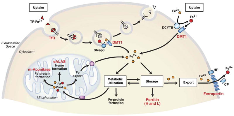
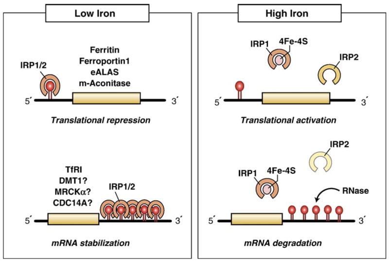
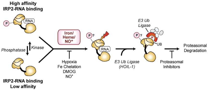
Both deficiencies and excesses of iron represent major public health problems throughout the world. Understanding the cellular and organismal processes controlling iron homeostasis is critical for identifying iron-related diseases and in advancing the clinical treatments for such disorders of iron metabolism. Iron regulatory proteins (IRPs) 1 and 2 are key regulators of vertebrate iron metabolism. These RNA binding proteins post-transcriptionally control the stability or translation of mRNAs encoding proteins involved in iron homeostasis thereby controlling the uptake, utilization, storage or export of iron. Recent evidence provides insight into how IRPs selectively control the translation or stability of target mRNAs, how IRP RNA binding activity is controlled by iron-dependent and iron-independent effectors, and the pathological consequences of dysregulation of the IRP system.


Figures

Fig. 1
Fig. 1
Control of mammalian iron homeostasis by the IRE/IRP regulatory network. A generic mammalian cell depicting the various roles of mammalian proteins encoded by IRE-containing mRNAs (red lettering). Tf-Fe3+ binds to Tf R on the cell surface where the TF-Fe3+/Tf R complex is endocytosed. Acidification of the endosome causes the release of Fe3+ (red balls) from TF where it is reduced to Fe2+ (yellow balls) by the ferrireductase Steap3 before export from the endosome by DMT1 (divalent metal transporter 1). ApoTf/Tf R complex is then returned to the cell surface where it dissociates and initiates another round of iron uptake. TF-mediated iron uptake is ubiquitous, but it is especially important in erythroid precursor cells where it is the primary source of iron for utilization in heme synthesis. DMT1 is also localized on the apical membrane of duodenal enterocytes where it transports Fe2+ reduced possibly by the membrane reductase DCYTB or a member of a Steap family of metalloreductases [300]. Iron taken up by the Tf R enters a cytosolic free iron pool thought to consist of Fe2+ bound to small molecular weight molecules. IRPs sense iron in this pool and regulate the translation of 5′ IRE-containing mRNAs (H- and L-ferritin subunits, eALAS (erythroid aminolevulinate synthase), m-aconitase, ferroportin) or the stability of 3′ IRE-containing mRNAs (Tf R and possibly DMT1). eALAS is a mitochondrial enzyme and the rate limiting enzyme in heme synthesis in precursor erythroid cells. Mitochondrial aconitase is an enzyme in the tricarboxylic acid cycle that requires a [4Fe–4S] cluster for activity. Iron that is not utilized or stored in ferritin is exported by ferroportin. Ferroportin is expressed in duodenal enterocytes, hepatocytes, placenta and macrophages. Export of iron is coupled to the oxidation of iron possibly by the membrane bound protein hephaestin (HP) or the serum multicopper oxidase ceruloplasmin (CP).
Fig. 2
Fig. 2
Cellular regulation of mammalian iron homeostasis by the IRPs. IRPs bind to IREs located in either the 5′- or 3′-UTRs of specific mRNAs. During low iron conditions, IRP1 and IRP2 bind with high affinity to 5′ IREs and to the five 3′ IREs in Tf R mRNA, resulting in the translational repression of 5′ IRE-containing mRNAs and the stabilization of the Tf R mRNA. During high iron conditions, IRPs lose their affinity for IREs, increasing translation of 5′ IRE-containing mRNAs and mediating degradation of the Tf R mRNA. Increased iron levels result in the conversion of the IRP1 RNA binding form into the [4Fe–4S] cluster c-acon form, while increased iron and/or heme levels mediate IRP2 proteasomal degradation. Whether the 3′ IRE in DMT1 mRNA is functional is not clear.
Fig. 3
Fig. 3
Predicted structure of ferritin and m-acon IRE regions. (A) The rat L-ferritin IRE plus flanking sequences (IRE region). Note that ferritin IRE is the central part of a large stem loop structure stabilized by sequences flanking the IRE. This structure has been confirmed in solution [143,150]. The 5′ and 3′ ends of the ferritin 28 nt IRE are indicated by arrows 1 and 2. The internal loop/bulge is indicated (bracket 3) as is the terminal loop (bracket 4). Potential basepairing between loop nucleotides 1 and 5 is shown. (B) m-acon 5′ UTR structure using the best example of an IRE-like fold obtained from computer predictions. In contrast to the ferritin and Fpn IRE regions, the m-acon IRE is not predicted to form as frequently. The highly conserved RNA sequence flanking [134] the m-acon IRE lacks extensive basepairing since, in contrast to ferritin and Fpn, the m-acon (and eALAS) IRE is <10 nt from the 5′ end. The 5′ CAP is shown for m-acon (★); ferritin cap is 28 nt from 5′ end of IRE. Structures were predicted by M-fold version 3.2.
Fig. 4
Fig. 4
Pathways for regulation of IRP1: IRP1 can be regulated through mechanisms dependent or independent of the [4Fe–4S] cluster. In the cluster dependent pathway IRP1 has dual roles either as a high affinity IRE RNA binding protein when devoid of the [4Fe–4S] cluster or it is the cytosolic isoform of aconitase (c-acon) when the [4Fe–4S] cluster is assembled. The c-acon form does not bind RNAwith high affinity. The [4Fe–4S] cluster is accessible to low molecular cluster perturbants including reactive oxygen species (ROS) such as O2 and H2O2 or reactive nitrogen species such as NO or ONOO. Hypoxia stabilizes the c-acon form by reducing the level of oxygen or ROS. IRP1 can be phosphorylated at S138 and S711; both sites are not accessible in the c-acon form but are readily phosphorylated in the RNA binding form. Studies with phosphomimetic mutants of IRP1 indicate that the Fe–S cluster can be assembled and the protein exhibits robust aconitase activity. However, the Fe–S cluster is much more sensitive to disruption by cluster perturbants including oxygen and hydrogen peroxide. Iron stimulates the degradation of S138 phosphomimetic mutants of IRP1 as well as S138 phosphorylated IRP1. The non-phosphorylated IRP1 apoprotein is also subject to iron induced protein degradation when the Fe–S cluster biogenesis pathway is impaired.
Fig. 5
Fig. 5
Proposed models for iron-dependent and iron-independent IRP2 regulation. Iron-dependent pathway: iron and/or heme transiently bind IRP2 modifying a specific amino acid(s), which is then recognized by a specific E3-ubiquitin ligase (HOIL-1) leading to IRP2 ubiquitination (UB) and proteasomal degradation. Alternatively, iron/oxygen activates a 2-OG-dioxygenase that hydroxylates IRP2, which provides a target site for a specific E3-ubiquitin ligase. NO+ stimulates IRP2 degradation while NOstabilizes IRP2. Hypoxia, iron chelators and the 2-OG-dioxygenase inhibitor DMOG also stabilize IRP2. Proteasome inhibitors block IRP2 degradation. IRP2 iron-independent pathway: phosphorylation regulates IRP2 RNA binding activity independent of iron. IRP2 can switch between a high-affinity phosphorylated form and a low-affinity dephosphorylated form, which are regulated by specific protein kinases and phosphatases. Both forms are substrates for iron-mediated degradation. IRP2 phosphorylation may allow IRP2 to alter iron-homeostasis independent of cellular iron concentration. IRP2 is displayed based on m-acon structure with three domains (I–III) connected by a linker to domain IV.

References

    1. Wood RJ, Ronnenberg AG. Iron. 10. Lippincott Williams and Wilkins; Philadelphia, PA: 2006.
    1. Lynch SR. The impact of iron fortification on nutritional anaemia. Best Pract Res Clin Haematol. 2005;18:333–346. - PubMed
    1. Hunt JM. Reversing productivity losses from iron deficiency: the economic case. J Nutr. 2002;132:794S–801S. - PubMed
    1. WHO. The World Health Report 2002-Reducing Risks, Promoting Health Life Styles. World Health Organization; 2002.
    1. Anonymous. Iron deficiency—United States, 1999–2000. MMWR. 2002;51:897–899. - PubMed

Publication types