

Brain atrophy in long-term abstinent alcoholics who demonstrate impairment on a simulated gambling task
- PMID: 16872844
- PMCID: PMC1868686
- DOI: 10.1016/j.neuroimage.2006.06.013
Brain atrophy in long-term abstinent alcoholics who demonstrate impairment on a simulated gambling task
Abstract
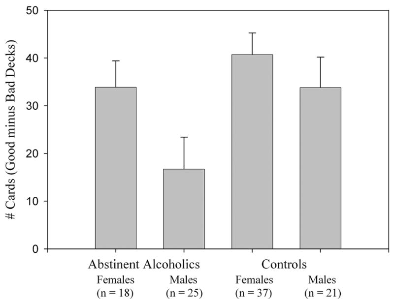
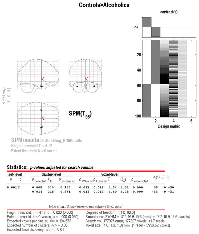
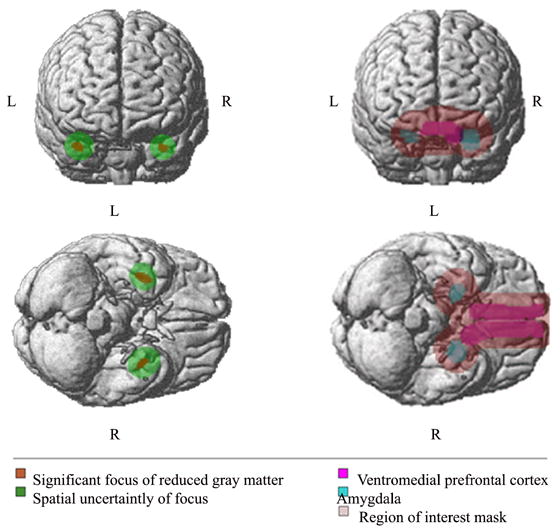
We recently demonstrated impairment on the Simulated Gambling Task (SGT) in long-term abstinent alcoholics (AbsAlc). Brain regions that have been shown to be necessary for intact SGT performance are the ventromedial prefrontal cortex (VMPFC) and the amygdala; patients with VMPFC or amygdalar damage demonstrate SGT impairments similar to those of substance abusing populations. We examined these brain regions, using T1-weighted MRIs, in the 101 participants from our previous study using voxel-based morphometry (VBM). VBM was performed using a modification we developed [Fein, G., Landman, B., Tran, H., Barakos, J., Moon, K., Di Sclafani, V., Shumway, R., 2006. Statistical parametric mapping of brain morphology: sensitivity is dramatically increased by using brain-extracted images as inputs. Neuroimage] of Baron's procedure, [], in which we use skull-stripped images as input. We also restricted the analysis to a ROI consisting of the amygdala and VMPFC as defined by the Talairach Daemon resource. Compared to the controls, the AbsAlc participants had significant foci of reduced gray matter density within the amygdala. Thus, SGT decision-making deficits are associated with reduced gray matter in the amygdala, a brain region previously implicated in similar decision-making impairments in neurological samples. This structurally based abnormality may be the result of long-term alcohol abuse or dependence, or it may reflect a pre-existing factor that predisposes one to severe alcoholism. From an image analysis perspective, this work demonstrates the increased sensitivity that results from using skull-stripped inputs and from restricting the analysis to a ROI. Without both of these methodological advances, no statistically significant finding would have been forthcoming from this work.
Figures
Similar articles
-
Impairment on a simulated gambling task in long-term abstinent alcoholics.Alcohol Clin Exp Res. 2004 Oct;28(10):1487-91. doi: 10.1097/01.alc.0000141642.39065.9b. Alcohol Clin Exp Res. 2004. PMID: 15597080 Free PMC article.
-
Normal performance on a simulated gambling task in treatment-naive alcohol-dependent individuals.Alcohol Clin Exp Res. 2006 Jun;30(6):959-66. doi: 10.1111/j.1530-0277.2006.00109.x. Alcohol Clin Exp Res. 2006. PMID: 16737453 Free PMC article.
-
Parietal gray matter volume loss is related to spatial processing deficits in long-term abstinent alcoholic men.Alcohol Clin Exp Res. 2009 Oct;33(10):1806-14. doi: 10.1111/j.1530-0277.2009.01019.x. Epub 2009 Jul 23. Alcohol Clin Exp Res. 2009. PMID: 19645730 Free PMC article.
-
Regional deficits in brain volume in schizophrenia: a meta-analysis of voxel-based morphometry studies.Am J Psychiatry. 2005 Dec;162(12):2233-45. doi: 10.1176/appi.ajp.162.12.2233. Am J Psychiatry. 2005. PMID: 16330585 Review.
-
[Decision-making and schizophrenia].Encephale. 2011 Dec;37 Suppl 2:S110-6. doi: 10.1016/S0013-7006(11)70036-7. Encephale. 2011. PMID: 22212839 Review. French.
Cited by
-
Daily marijuana use is not associated with brain morphometric measures in adolescents or adults.J Neurosci. 2015 Jan 28;35(4):1505-12. doi: 10.1523/JNEUROSCI.2946-14.2015. J Neurosci. 2015. PMID: 25632127 Free PMC article.
-
Common Gray Matter Reductions in Alcohol Use and Obsessive-Compulsive Disorders: A Meta-analysis.Biol Psychiatry Glob Open Sci. 2021 Nov 27;2(4):421-431. doi: 10.1016/j.bpsgos.2021.11.010. eCollection 2022 Oct. Biol Psychiatry Glob Open Sci. 2021. PMID: 36324652 Free PMC article.
-
Aberrant paralimbic gray matter in criminal psychopathy.J Abnorm Psychol. 2012 Aug;121(3):649-58. doi: 10.1037/a0026371. Epub 2011 Dec 12. J Abnorm Psychol. 2012. PMID: 22149911 Free PMC article.
-
Neuronal nicotinic acetylcholine receptors as pharmacotherapeutic targets for the treatment of alcohol use disorders.CNS Neurol Disord Drug Targets. 2010 Mar;9(1):60-76. doi: 10.2174/187152710790966597. CNS Neurol Disord Drug Targets. 2010. PMID: 20201817 Free PMC article. Review.
-
Impulsive-antisocial psychopathic traits linked to increased volume and functional connectivity within prefrontal cortex.Soc Cogn Affect Neurosci. 2017 Jul 1;12(7):1169-1178. doi: 10.1093/scan/nsx042. Soc Cogn Affect Neurosci. 2017. PMID: 28402565 Free PMC article.
References
-
- Aggleton J, Mishkin M. The amygdala: Sensory gateway to the emotions. In: Plutchik R, Kellerman H, editors. Emotional theory, research, and experience. Vol. 3. Academic Press; New York: 1986. pp. 281–299.
-
- Ashburner J, Friston KJ. Voxel-based morphometry--the methods. Neuroimage. 2000;11(6 Pt 1):805–21. - PubMed
-
- Bar-On R, Tranel D, Denburg NL, Bechara A. Exploring the neurological substrate of emotional and social intelligence. Brain. 2003;126(Pt 8):1790–800. - PubMed
-
- Baron JC, Chetelat G, Desgranges B, Perchey G, Landeau B, de la Sayette V, Eustache F. In vivo mapping of gray matter loss with voxel-based morphometry in mild Alzheimer’s disease. Neuroimage. 2001;14(2):298–309. - PubMed
-
- Baxter MG, Murray EA. The amygdala and reward. Nat Rev Neurosci. 2002;3(7):563–73. - PubMed
Publication types
MeSH terms
Grants and funding
LinkOut - more resources
Full Text Sources
Medical
