. 2007 Jan;7(1-2):131-6.
doi: 10.1016/j.modgep.2006.06.002. Epub 2006 Jun 14.

Ubl4b, an X-derived retrogene, is specifically expressed in post-meiotic germ cells in mammals

Affiliations

Ubl4b, an X-derived retrogene, is specifically expressed in post-meiotic germ cells in mammals

Fang Yang et al. Gene Expr Patterns. 2007 Jan.

Abstract

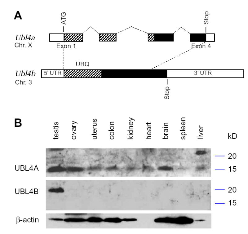
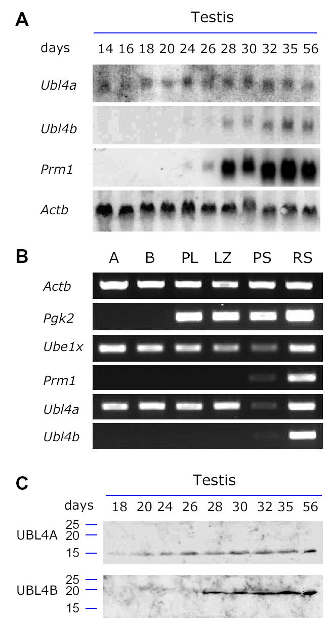
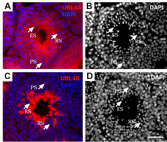
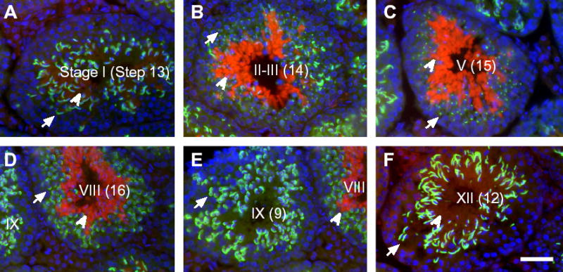
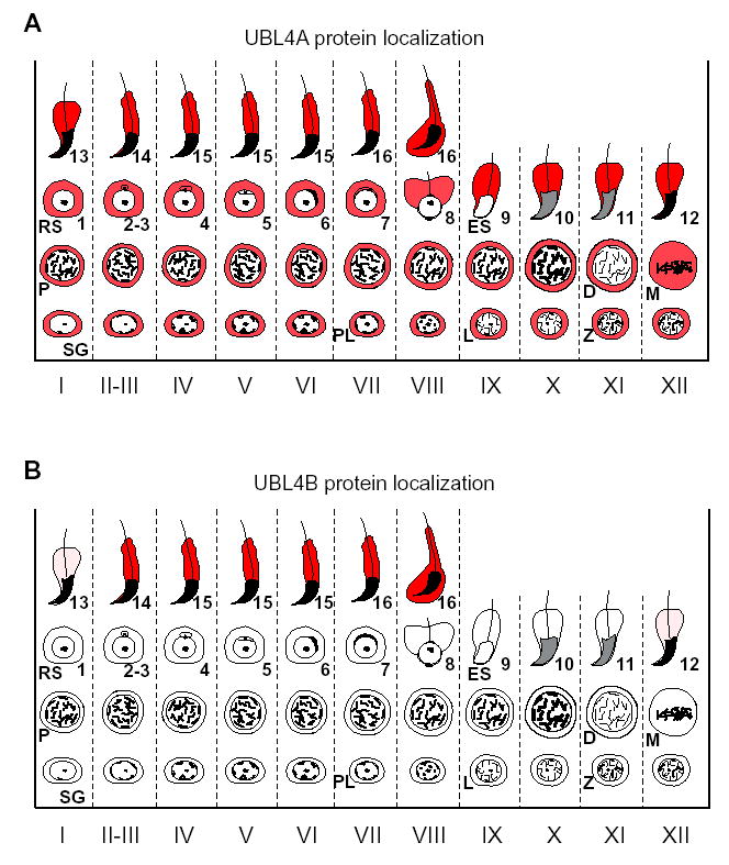
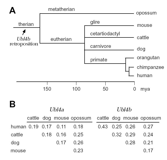
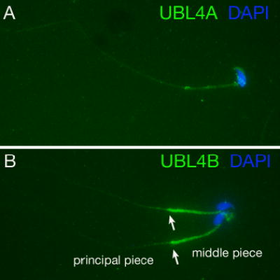
Post-translational modification by ubiquitin and ubiquitin-related proteins plays critical roles in protein degradation and in regulation of essential cellular processes. In mammals, transcription grinds to a halt during late spermiogenesis due to compaction of the spermatid genome, which creates a special need for robust post-transcriptional regulation. Here, we report the finding of a novel mouse ubiquitin-like protein, UBL4B. Ubl4b is a testis-specific autosomal gene. Ubl4b lacks introns and evidently arose from an X-linked intron-bearing housekeeping gene, Ubl4a, by retroposition during mammalian evolution. While Ubl4a is expressed throughout spermatogenesis, Ubl4b is restricted to post-meiotic germ cells. Ubl4a is highly conserved, but Ubl4b has undergone rapid evolution and may have evolved new functions. Our data suggest that evolution of Ubl4b is not due to meiotic sex chromosome inactivation (MSCI). Alternatively, origination of Ubl4b was due to MSCI, but Ubl4b eventually evolved to be restricted to post-meiotic germ cells.


Figures

Fig. 1
Fig. 1
Ubl4b is a testis-specific retrogene. (A) Comparison of the exon/intron structures of mouse Ubl4a and Ubl4b. Exons 1 and 4 of Ubl4a are labeled. Introns and exons are drawn to scale. Coding region is shown in hatch pattern and black. The hatched region indicates the ubiquitin (UBQ) domain. There is little sequence identity between Ubl4a and Ubl4b at the nucleotide level. (B) Western blot analysis of UBL4A and UBL4B in mouse tissues. Bacterially expressed recombinant UBL4A and UBL4B were specifically recognized with respective antibodies (data not shown). Western blotting with pre-immune sera was negative (data not shown). Molecular weight markers are shown in kD.
Fig. 2
Fig. 2
Differential expression of UBL4A and UBL4B during spermatogenesis. Postnatal juvenile testes were collected. The number of days after birth was indicated. (A) Northern blot analysis. ATTT serves as a ubiquitous gene control. Prm1 is a post-meiotically expressed germ cell-specific gene. (B) RT-PCR expression analysis using germ cell preparations. Relative levels of transcripts among different germ cell types were assayed by RT-PCR. Four control genes were used as previously described (Wang, et al., 2005). In addition to Actb and Prm1, Pgk2 initiates transcription at the onset of meiosis. Ube1x is a control for undergoing MSCI as previously documented (Odorisio, et al., 1996). A: type A spermatogonia; B: type B spermatogonia; PL: preleptotene spermatocytes; LZ: leptotene plus zygotene spermatocytes; PS: adult pachytene spermatocytes; RS: round spermatids. (C) Western blot analysis. 30 μg of testicular protein extracts for each sample was used. Molecular weight standards were marked in kD.
Fig. 3
Fig. 3
Subcellular localization of UBL4A and UBL4B in male germ cells. Adult mouse testis sections were immunostained with anti-UBL4A or anti-UBL4B antibodies (both shown in red). Nuclear DNA was stained with DAPI. (A) UBL4A is distributed in the cytoplasm of all germ cells. (C) Localization of UBL4B is restricted to elongating spermatids. Panels B and D are identical to Panels A and C, respectively, except that nuclei staining alone is shown to distinguish among different testicular cell types. PS, pachytene spermatocytes; RS, round spermatids; ES, elongating spermatids. Scale bar, 50 μm
Fig. 4
Fig. 4
Stage-dependent distribution of UBL4B in mouse spermiogenesis. (A-F) Expression of UBL4B in representative tubules. Testis sections were immunostained with anti-UBL4B antibodies (red, indicated by arrow heads), anti-ACRV1 (green, indicated by arrows), and DAPI (blue). Anti-UBL4B antiserum did not show cross reactivity with UBL4A (Fig. 1C). The morphology of acrosomes (stained by anti-ACRV1 antibodies) and nuclei (stained by DAPI) was used to determine the stages of seminiferous tubules. The stage number is shown in roman numerals. The Arabic number in parenthesis designates the step of most advanced spermatids. Elongated spermatids are multiple layers of germ cells located in the lumen of seminiferous tubules. (D, E) Comparison of two adjacent tubules at Stages VIII and IX. As Step 16 spermatozoa are released from the seminiferous epithelium in Stage VIII, UBL4B is not observed in the subsequent Stage IX tubules. Scale bar, 50 μm.
Fig. 5
Fig. 5
Summary of UBL4A and UBL4B protein localization during spermatogenesis. Expression of UBL4A and UBL4B protein is shown in red. Stages (I-XII) of spermatogenesis are shown at the bottom. Representative types of germ cells are indicated. SG, spermatogonia; PL, preleptotene spermatocytes; L, leptotene; Z, zygotene; P, pachytene; D, diplotene; M, metaphase spermatocyte; RS, round spermatid; ES, elongating spermatid.
Fig. 6
Fig. 6
Evolution of the Ubl4b retrogene. (A) Timing of the Ubl4b retroposition. The timescale was drawn as described previously (mya, million years ago) (Kumar and Hedges, 1998). By searching the genomic sequence databases, we identified the Ubl4b orthologues in opossum, mouse, cattle, dog, orangutan, and human. (B) Ka/Ks ratios calculated from pairwise comparisons in five species. Calculation was performed for Ubl4a and Ubl4b separately. The cDNA sequences of Ubl4a and Ubl4b orthologues were retrieved from GenBank. The opossum Ubl4a cDNA sequence was electronically predicted from genomic sequences generated by Broad Institute (Cambridge, MA, USA). The coding sequences were aligned using CLUSTAL W (Jeanmougin, et al., 1998). The Ka/Ks ratios were calculated according to Li’s method as implemented in the GCG software (Li, 1993).
Supplementary Figure S1
Localization of UBL4A and UBL4B in epididymal sperm. Sperm from epididymis were fixed in 4% PFA and were immunostained with affinity-purified anti-UBL4A (green) or anti-UBL4B antibodies (green) and DAPI (blue). (A) Anti-UBL4A antibodies weakly stained both sperm tail and periphery of sperm head. (B) Anti-UBL4B antibodies produced a strong staining of the middle piece and a relatively weak staining of the principal piece. Two sperm were shown in (B). The arrows demarcate the possible junction between the middle piece and principal piece.

References

    1. Bebington C, Doherty FJ, Fleming SD. The possible biological and reproductive functions of ubiquitin. Hum Reprod Update. 2001;7:102–111. - PubMed
    1. Bradley J, Baltus A, Skaletsky H, Royce-Tolland M, Dewar K, Page DC. An X-to-autosome retrogene is required for spermatogenesis in mice. Nat Genet. 2004;36:872–876. - PubMed
    1. Dahl HH, Brown RM, Hutchison WM, Maragos C, Brown GK. A testis-specific form of the human pyruvate dehydrogenase E1 alpha subunit is coded for by an intronless gene on chromosome 4. Genomics. 1990;8:225–232. - PubMed
    1. Dass B, McMahon KW, Jenkins NA, Gilbert DJ, Copeland NG, MacDonald CC. The gene for a variant form of the polyadenylation protein CstF-64 is on chromosome 19 and is expressed in pachytene spermatocytes in mice. J Biol Chem. 2001;276:8044–8050. - PubMed
    1. Elliott DJ, Venables JP, Newton CS, Lawson D, Boyle S, Eperon IC, Cooke HJ. An evolutionarily conserved germ cell-specific hnRNP is encoded by a retrotransposed gene. Hum Mol Genet. 2000;9:2117–2124. - PubMed

Publication types