Review
. 2006 Aug 29;361(1472):1351-64.
doi: 10.1098/rstb.2006.1874.

Proton-coupled electron transfer: the mechanistic underpinning for radical transport and catalysis in biology

Affiliations
Review

Proton-coupled electron transfer: the mechanistic underpinning for radical transport and catalysis in biology

Steven Y Reece et al. Philos Trans R Soc Lond B Biol Sci. .

Abstract

Charge transport and catalysis in enzymes often rely on amino acid radicals as intermediates. The generation and transport of these radicals are synonymous with proton-coupled electron transfer (PCET), which intrinsically is a quantum mechanical effect as both the electron and proton tunnel. The caveat to PCET is that proton transfer (PT) is fundamentally limited to short distances relative to electron transfer (ET). This predicament is resolved in biology by the evolution of enzymes to control PT and ET coordinates on highly different length scales. In doing so, the enzyme imparts exquisite thermodynamic and kinetic controls over radical transport and radical-based catalysis at cofactor active sites. This discussion will present model systems containing orthogonal ET and PT pathways, thereby allowing the proton and electron tunnelling events to be disentangled. Against this mechanistic backdrop, PCET catalysis of oxygen-oxygen bond activation by mono-oxygenases is captured at biomimetic porphyrin redox platforms. The discussion concludes with the case study of radical-based quantum catalysis in a natural biological enzyme, class I Escherichia coli ribonucleotide reductase. Studies are presented that show the enzyme utilizes both collinear and orthogonal PCET to transport charge from an assembled diiron-tyrosyl radical cofactor to the active site over 35A away via an amino acid radical-hopping pathway spanning two protein subunits.


Figures

Figure 1
Figure 1
Two basic scenarios for PCET in biology: collinear PCET, which may involve bond making/breaking; and orthogonal PCET, where PT to a base (B:) occurs along a separate coordinate than ET. This pathway is usually accompanied by bond breaking/making.
Figure 2
Figure 2
Square scheme describing the thermochemistry of different pathways of a HAT reaction. The overall reaction free energy is obtained by combining the relevant reduction potentials and pKa values corresponding to stepwise ET/PT (or PT/ET) reactions around the edges. Yet, in many cases, direct HAT along the diagonal is favoured in order to avoid energetic intermediates.
Figure 3
Figure 3
The 3.4 Å resolution structure of the oxygen evolving complex (OEC) and the immediate peptide environment adapted from Ferreira et al. (2004). The direction of proposed PT and ET pathways are indicated with arrows.
Figure 4
Figure 4
(a) Model collinear PCET complexes assembled from a symmetric carboxylic acid dimer interface or an asymmetric amidinium–carboxylate interface. D is a photo-excitable donor; A is an electron acceptor. (b) An assembly used to study the role of the mediating proton in collinear PCET by the temperature dependence of the deuterium isotope effect (Damrauer et al. 2004; Hodgkiss et al. 2006).
Figure 5
Figure 5
Temperature dependence of the rate of PCET in the porphyrin assembly shown in figure 4b for a protiated (solid circles) and deuterated (open circles) amidinium–carboxylate interface in the solvent 2-methyl tetrahydrofuran (2-MeTHF). Data are presented in a modified Arrhenius form with linear fits (Hodgkiss et al. 2006). Reproduced with permission from J. Phys. Chem. B. Copyright © 2006, Am. Chem. Soc.
Figure 6
Figure 6
(a) Schematic of model systems for studying fixed-distance orthogonal PCET. Excitation of the photo-oxidant triggers ET from the iron porphyrin, which donates a proton to the base (B) appended to the Hangman pillar. The key features of this design are that the ET and PT kinetics can be tuned through variation of the spacer and pillar, respectively, and driving forces for each of these processes can be tuned independently by varying the photo-oxidant and the base. Alternatively, if a photo-excitable donor is appended to the porphyrin, the PCET reaction can be run in the opposite direction (ET to the porphyrin along with protonation upon reduction). (b) A line drawing and crystal structure of the Hangman porphyrin showing the water channel adapted from (Yeh et al. 2001a).
Figure 7
Figure 7
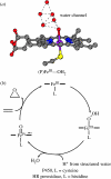
(a) High-resolution structure of cytochrome P450, displaying a water channel above the haem adapted from Poulos et al. (1986). (b) The peroxo-shunt mechanism of mono-oxygenases produces compound I ((P·+)FeIV=O), which oxidizes substrates by their nucleophilic attack on the electrophilic oxo of the (P·+)FeIV=O core. Reproduced with permission from Inorg. Chem. 44, 6879–6892. Copyright © 2006, Am. Chem. Soc.
Figure 8
Figure 8
Conserved residues of class I RNR that compose the putative PCET pathway for radical transport from ·Y122 in R2 to C439 in the R1 active site (Seyedsayamdost et al. 2005b). Distances are from the separate crystal structures of the R1 (Uhlin & Eklund 1994) and R2 (Högbom et al. 2003) subunit from the E. coli enzyme. Reproduced with permission from J. Am. Chem. Soc. (in press). Copyright © 2006, Am. Chem. Soc.
Figure 9
Figure 9
Redox potential regimes of RNR activity (Seyedsayamdost et al. 2005b). Relative activities of FnY356-R2s versus Y-R2, plotted as a function of peak reduction potential difference between the corresponding Ac-FnY·-NH2 and Ac-Y·-NH2: (blue filled and open circles) 3,5-F2Y356-R2, (red filled and open circles) 2,3-F2Y356-R2, (magenta circles) 2,3,5-F3Y356-R2, (green filled and open circles) 2,3,6-F3Y356-R2, and (orange circles) F4Y356-R2. Filled circles represent data points where pH<pKa of the corresponding Ac-FnY-NH2; open circles represent data points where pH>pKa of the corresponding Ac-FnY-NH2. The three different regimes of RNR activity are highlighted as either gated by a physical/conformational change (regime 1), rate-limited by radical transport (regime 2), or reduced to background levels (regime 3) depending on the peak reduction potential difference between the corresponding Ac-FnY·-NH2 and Ac-Y·-NH2. Reproduced with permission from J. Am. Chem. Soc. (in press). Copyright © 2006, Am. Chem. Soc.
Figure 10
Figure 10
Experimental design for studying the kinetics of radical transport along ·Y356→Y731→Y730→C439 pathway. ·Y356 is generated photochemically by a proximal photo-oxidant (red circle) on the R2C19 peptide. NDP, nucleoside diphosphate substrate; dNDP, deoxynucleoside diphosphate product; R2C19, 19-mer C-terminal peptide tail of R2.
Figure 11
Figure 11
Light-initiated single turnover experiments in R1-peptide complexes. Samples were irradiated using 299 nm long pass filters at room temperature for 2 min. Light (cyan) and dark (grey) bars correspond to light reactions and dark controls, respectively. R1 was purified and dC product quantitated as previously described (Chang et al. 2004c).
Figure 12
Figure 12
Proposed model for radical transport in RNR. The mode of transport at the interface (between Y356 and 731) is undefined.
Figure 13
Figure 13
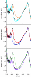
The transient absorption spectrum of (black) BPA-Y-OMe, (cyan) BPA-3-FY-OMe, (blue) BPA-3,5-F2Y-OMe, (red) BPA-2,3-F2Y-OMe, (green) BPA-2,3,6-F3Y-OMe, (violet) BPA-2,3,5-F3Y-OMe and (orange) BPA-F4Y-OMe obtained 100 ns after excitation of ca 500 μM solutions of each dipeptide buffered to pH 4.0 with 20 mM succinic acid and normalized to the ketyl radical absorption peak at 547 nm (Seyedsayamdost et al. 2006a). Reproduced with permission from J. Am. Chem. Soc. Copyright © 2006, Am. Chem. Soc.

Similar articles

Cited by

References

    1. Aubert C, Vos M.H, Mathis P, Eker A.P.M, Brettel K. Intraprotein radical transfer during photoactivation of DNA photolyase. Nature. 2000;405:586–590. doi:10.1038/35014644 - DOI - PubMed
    1. Baldwin J, Krebs C, Ley B.A, Edmondson D.E, Huynh B.H, Bollinger J.M., Jr Mechanism of rapid electron transfer during oxygen activation in the R2 subunit of Escherichia coli ribonucleotide reductase. 1. Evidence for a transient tryptophan radical. J. Am. Chem. Soc. 2000;122:12 195–12 206. doi:10.1021/ja001278u - DOI
    1. Barber J, Ferreira K, Maghlaoui K, Iwata S. Structural model of the oxygen-evolving center of photosystem II with mechanistic implications. Phys. Chem. Chem. Phys. 2004;6:4737–4742. doi:10.1039/b407981g - DOI
    1. Barry B, Babcock G.T. Tyrosine radicals are involved in the photosynthetic oxygen-evolving system. Proc. Natl Acad. Sci. USA. 1987;84:7099–7103. doi:10.1073/pnas.84.20.7099 - DOI - PMC - PubMed
    1. Beratan D.N, Onuchic J.N, Hopfield J.J. Electron tunneling through covalent and noncovalent pathways in proteins. J. Chem. Phys. 1987;86:4488–4498. doi:10.1063/1.452723 - DOI

LinkOut - more resources