. 2006 Aug;80(16):7816-31.
doi: 10.1128/JVI.00532-06.

Cleavage map and proteolytic processing of the murine norovirus nonstructural polyprotein in infected cells

Affiliations

Cleavage map and proteolytic processing of the murine norovirus nonstructural polyprotein in infected cells

Stanislav V Sosnovtsev et al. J Virol. 2006 Aug.

Abstract

Murine norovirus (MNV) is presently the only member of the genus Norovirus in the Caliciviridae that can be propagated in cell culture. The goal of this study was to elucidate the proteolytic processing strategy of MNV during an authentic replication cycle in cells. A proteolytic cleavage map of the ORF1 polyprotein was generated, and the virus-encoded 3C-like (3CL) proteinase (Pro) mediated cleavage at five dipeptide cleavage sites, 341E/G342, Q705/N706, 870E/G871, 994E/A995, and 1177Q/G1178, that defined the borders of six proteins with the gene order p38.3 (Nterm)-p39.6 (NTPase)-p18.6-p14.3 (VPg)-p19.2 (Pro)-p57.5 (Pol). Bacterially expressed MNV 3CL Pro was sufficient to mediate trans cleavage of the ORF1 polyprotein containing the mutagenized Pro sequence into products identical to those observed during cotranslational processing of the authentic ORF1 polyprotein in vitro and to those observed in MNV-infected cells. Immunoprecipitation and Western blot analysis of proteins produced in virus-infected cells demonstrated efficient cleavage of the proteinase-polymerase precursor. Evidence for additional processing of the Nterm protein in MNV-infected cells by caspase 3 was obtained, and Nterm sequences 118DRPD121 and 128DAMD131 were mapped as caspase 3 cleavage sites by site-directed mutagenesis. The availability of the MNV nonstructural polyprotein cleavage map in concert with a permissive cell culture system should facilitate studies of norovirus replication.


Figures
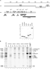
FIG. 1.
FIG. 1.
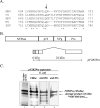
Expression of MNV polyprotein fragments in E. coli and specificities of the corresponding antisera. (A) Schematic diagram showing the organization of the MNV genome, putative cleavage products of the virus ORF1 polyprotein, and location of cDNA fragments expressed in E. coli. Predicted dipeptide cleavage sites are indicated with arrows, and the calculated molecular weights (MW) (in thousands) of the expected cleavage products are shown. The cDNA fragments engineered for expression in E. coli are depicted as dark gray bars. Fusion proteins corresponding to the selected ORF1 regions were purified using Ni-NTA agarose. For lanes 1 to 6, a sample of each region-specific protein (identified above the lanes) was analyzed by SDS-PAGE followed by Coomassie blue staining. (B) Immunoprecipitation of the MNV ORF1 nonstructural proteins synthesized in a TNT reaction from the genomic full-length clone p20.3 using a panel of region-specific antisera. Radiolabeled MNV ORF1 polyprotein cleavage products (lane 1) were immunoprecipitated with either pre- or postimmunization sera raised against Nterm protein (lanes 2 and 3), p18 protein (lanes 4 and 5), VPg protein (lanes 6 and 7), Pro protein (lanes 8 and 9), and ProPol protein (lanes 10 and 11) and subjected to SDS-PAGE in a 12% Tris-glycine gel followed by autoradiography of the dried gel. Lane 12, TNT products derived from the MNV ORF1 clone pDNORF1 in which the first two AUG codons were abolished. Proteins that were not identified in this study and that likely represented nonspecific products in the TNT reaction are indicated with asterisks.
FIG. 2.
FIG. 2.
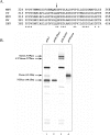
Mapping of VPg, 3CL Pro, and polymerase. (A) Schematic representation of the clones used to express VPg-, Pro-, and Pol-containing ORF1 polyprotein sequences in bacteria. Observed molecular masses of the detected cleavage products used in the direct N-terminal protein sequence analysis are shown at the top of the diagram, and identified cleavage sites are shown at the bottom of the diagram. (B) SDS-PAGE and Western blot analysis of the MNV-specific proteins encoded in plasmids pMBN and pMVP and expressed in bacteria. Bacterial cells carrying either the pMBN or pMVP plasmid or the pET-28a vector plasmid were induced with IPTG, and lysates of the cells were prepared as described in Materials and Methods. Aliquots of the bacterial cell lysates were subjected to SDS-PAGE and visualized with Coomassie blue stain. Lanes 1, 3, and 5, uninduced cells harboring pET-28a, pMBN, and pMVP, respectively; lanes 2, 4, and 6, induced cells harboring the corresponding plasmids. Expressed recombinant proteins are indicated with arrows. To confirm the identity of the 16-kDa protein as VPg and the 19-kDa protein as proteinase, Western blot analysis using anti-VPg and anti-Pro sera was performed on lysates of induced bacterial cells harboring pET-28a (lane 7), pMBN (lane 8), and pMVP (lane 9). N-terminal sequence analysis of the VPg protein was performed using a protein that was SDS-PAGE purified from the lysate of IPTG-induced cells harboring pMBN. His6-tagged full-length polymerase (Pol-His) and proteinase (Pro-His) proteins were purified from soluble fractions of cells carrying pMBN and pMVP, respectively, for direct N-terminal sequence analysis using IMAC chromatography. MW, molecular weight (in thousands). (C) Profile of radioactivity released during Edman degradation of the [35S]methionine-labeled Pol-His protein derived in vitro from pMBN. Coupled transcription and translation of pMBN were performed in the TNT system in the presence of [35S]methionine. The 58-kDa protein (Pol-His) was electrotransferred onto a polyvinylidene difluoride membrane following SDS-PAGE and subjected to microsequencing. The released radioactivity was determined for each cycle and is plotted against the residue number. The predicted N-terminal sequence of Pol-His is shown below the histogram, and the mapped cleavage site is indicated with arrow. The first amino acid residue of Pol-His produced in bacteria is denoted with a circle.
FIG. 3.
FIG. 3.
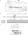
Analysis of the cleavage site between NTPase and p18. (A) Alignment of the amino acid sequence of the putative junction of the MNV NTPase and p18 proteins with those of CV, MDV, NV, and SHV. Numbers correspond to the ORF1 polyprotein sequence of each virus, and asterisks indicate conserved amino acids. The cleavage site in this region, identified previously for human norovirus strains, is indicated with an arrow. (B) Schematic representation of the pF2R2Pro plasmid that was engineered to contain the MNV-1 NTPase-p18 junction and an active 3CL Pro. The chimeric protein was fused to the vector sequence (gray box) at its N terminus, and the entire VPg and p18 C-terminal sequences were deleted. (C) SDS-PAGE analysis of proteins encoded in pF2R2Pro when expressed in vitro and in E. coli. Lane 1, in vitro TNT translation products derived from pF2R2Pro (autoradiography); lane 2, IMAC-purified proteins isolated from the insoluble fraction of the induced bacterial cells carrying pF2R2Pro stained with Coomassie blue (Cblue); lanes 3 and 4, Western blot analysis of the same protein sample using PentaHis antibody (QIAGEN) and anti-Pro serum, respectively. The two major pF2R2Pro proteins were expressed in both in vitro translation reactions and bacteria. The N-terminal sequence of the 24-kDa protein (NKVYDFDAG) is shown.
FIG. 4.
FIG. 4.
Analysis of the cleavage site between Nterm and NTPase. (A) Alignment of the amino acid sequence of the putative junction of the MNV Nterm and NTPase proteins with those of CV, MDV, NV, and SHV. Numbers correspond to the ORF1 polyprotein sequence of each virus, and asterisks indicate conserved amino acids. (B) Comparative analysis of the MNV ORF1 polyprotein processing products synthesized in vitro from full-length genomic clone p20.3 (lane 1) and its derivative, p20.3m341, carrying an Nterm-NTPase cleavage site mutation (341EG→341AG) (lane 3). Lanes 2 and 4, in vitro-radiolabeled proteins derived from clones pCINterm and pCINTPase, encoding predicted sequences of Nterm (aa 1 to 341) and NTPase (aa 342 to 705), respectively.
FIG. 5.
FIG. 5.
Expression of MNV-1 proteins in infected cells. (A to C and E to H) Virus proteins in MNV-1-infected cells collected at different time points following infection were analyzed. Ten micrograms of each cell lysate prepared from collected cells was resolved on a 4 to 20% polyacrylamide gel by SDS-PAGE and transferred electrophoretically onto a nitrocellulose membrane. The membrane was probed with serum collected from a mouse that had undergone infection with MNV-1 (A). The same lysates from the MNV-infected cells were probed with region-specific antisera: anti-capsid (B), anti-Nterm (C), anti-ProPol (E), anti-Pro (F), anti-p18 (G), and anti-VPg (H) sera. To compare the mobilities of the virus proteins synthesized in infected cells and in vitro, TNT products derived from pCIORF2, pCIPol, pCIPro, and pCINterm were analyzed concurrently with the corresponding cell lysate samples probed with anti-capsid (B, lane 8), anti-Nterm (C, lane 8), and anti-ProPol (E, lane 8) sera. (D) Comparison of MNV Nterm proteins expressed in vitro and in infected cells. Immunoprecipitation (IP) of the radiolabeled Nterm protein synthesized in the in vitro system from pCINterm and p20.3 (lanes 1 and 3, respectively) and in infected cells (lane 2) is shown. Lane 4, the MNV ORF1 nonstructural proteins derived from p20.3 in vitro. (I) Comparative immunoprecipitation analysis of p18-, VPg-, and Pro-related proteins derived by in vitro cleavage of the ORF1 polyprotein or synthesized in MNV-infected cells. The radiolabeled p18-related proteins were precipitated using anti-p18 serum from the ORF1 (lane 1) and pCIP18 (lane 3) translation mixture or from MNV-infected cells (lane 2). The radiolabeled VPg-related proteins were precipitated using anti-VPg serum from the ORF1 translation mixture (lane 4) or from MNV-infected cells (lane 5). The radiolabeled Pro-related proteins were precipitated using anti-Pro serum from the ORF1 (lane 6) and pCIPro (lane 8) translation mixture or from MNV-infected cells (lane 7). (J) Expression of the p18 protein in in vitro systems. Lanes 1 and 2, immunoprecipitation of the p18 protein derived from pCIP18 using the TNT T7 Coupled Reticulocyte Lysate system (RR) or the TNT T7 Coupled Wheat Germ Extract system (WGE) (Promega), respectively; lane 3, the MNV ORF1 nonstructural proteins derived from p20.3 using the TNT T7 Coupled Reticulocyte Lysate system. Modified and free forms of the p18 protein are indicated with arrows. MW, molecular weight (in thousands).
FIG. 5.
FIG. 5.
Expression of MNV-1 proteins in infected cells. (A to C and E to H) Virus proteins in MNV-1-infected cells collected at different time points following infection were analyzed. Ten micrograms of each cell lysate prepared from collected cells was resolved on a 4 to 20% polyacrylamide gel by SDS-PAGE and transferred electrophoretically onto a nitrocellulose membrane. The membrane was probed with serum collected from a mouse that had undergone infection with MNV-1 (A). The same lysates from the MNV-infected cells were probed with region-specific antisera: anti-capsid (B), anti-Nterm (C), anti-ProPol (E), anti-Pro (F), anti-p18 (G), and anti-VPg (H) sera. To compare the mobilities of the virus proteins synthesized in infected cells and in vitro, TNT products derived from pCIORF2, pCIPol, pCIPro, and pCINterm were analyzed concurrently with the corresponding cell lysate samples probed with anti-capsid (B, lane 8), anti-Nterm (C, lane 8), and anti-ProPol (E, lane 8) sera. (D) Comparison of MNV Nterm proteins expressed in vitro and in infected cells. Immunoprecipitation (IP) of the radiolabeled Nterm protein synthesized in the in vitro system from pCINterm and p20.3 (lanes 1 and 3, respectively) and in infected cells (lane 2) is shown. Lane 4, the MNV ORF1 nonstructural proteins derived from p20.3 in vitro. (I) Comparative immunoprecipitation analysis of p18-, VPg-, and Pro-related proteins derived by in vitro cleavage of the ORF1 polyprotein or synthesized in MNV-infected cells. The radiolabeled p18-related proteins were precipitated using anti-p18 serum from the ORF1 (lane 1) and pCIP18 (lane 3) translation mixture or from MNV-infected cells (lane 2). The radiolabeled VPg-related proteins were precipitated using anti-VPg serum from the ORF1 translation mixture (lane 4) or from MNV-infected cells (lane 5). The radiolabeled Pro-related proteins were precipitated using anti-Pro serum from the ORF1 (lane 6) and pCIPro (lane 8) translation mixture or from MNV-infected cells (lane 7). (J) Expression of the p18 protein in in vitro systems. Lanes 1 and 2, immunoprecipitation of the p18 protein derived from pCIP18 using the TNT T7 Coupled Reticulocyte Lysate system (RR) or the TNT T7 Coupled Wheat Germ Extract system (WGE) (Promega), respectively; lane 3, the MNV ORF1 nonstructural proteins derived from p20.3 using the TNT T7 Coupled Reticulocyte Lysate system. Modified and free forms of the p18 protein are indicated with arrows. MW, molecular weight (in thousands).
FIG. 6.
FIG. 6.
Detection and mapping of the MNV Nterm protein cleavage by caspase 3. (A) Comparison of cleavage products of the MNV Nterm protein in virus-infected cells with those generated by cleavage with the virus Pro protein and caspase 3 in vitro. Lanes 1 and 2, immunoprecipitation of the radiolabeled Nterm protein from the virus-infected cells using pre- and postimmunization anti-Nterm serum, respectively; lane 4, products of incubation of the radiolabeled Nterm protein derived by TNT translation from pCINterm (Nterm TNT) (2 μl of the translational mixture) with recombinant virus proteinase (HisPro) (2 μg, 3 h, 37°C), respectively (the 38-kDa cleavage product is indicated with an arrow); lanes 6 to 10, products of incubation of the Nterm TNT with lysates (10 μl) of the cell collected at different times of infection; lane 11, incubation of the Nterm TNT with one unit of mouse recombinant caspase 3 (Cas3). Untreated Nterm TNT (incubated 3 h at 37°C) was included as a control (lanes 3 and 5). Asterisks denote cleavage products of the Nterm protein precipitated from infected cells that had mobilities similar to those generated by caspase 3 cleavage. MW, molecular weight (in thousands). (B) Schematic representation of the Nterm clones pCINtermG103, pCINtermG121, and pCINtermG131 derived from pCINterm by site-directed mutagenesis of the caspase 3 putative cleavage sites 100DMSD103, 118DRPD121, and 128DAMD131, respectively. Predicted caspase 3 cleavage products of the mutant Nterm proteins are depicted as gray boxes, and their molecular masses are shown above the diagram. (C) SDS-PAGE analysis of cleavage products obtained after incubation (3 h at 37°C) of caspase 3 (Cas3) with TNT-radiolabeled Nterm (lane 4) or with its mutant versions, NtermG103 (lane 5), NtermG121 (lane 6), and NtermG131 (lane 7). TNT mixture without plasmid (lane 1), TNT-derived Nterm protein (lane 2), and the same protein after 3 h of incubation in assay buffer (lane 3) were included as controls.

Similar articles

Cited by

References

    1. Al-Molawi, N., V. A. Beardmore, M. J. Carter, G. E. Kass, and L. O. Roberts. 2003. Caspase-mediated cleavage of the feline calicivirus capsid protein. J Gen. Virol. 84:1237-1244. - PubMed
    1. Ando, T., J. S. Noel, and R. L. Fankhauser. 2000. Genetic classification of “Norwalk-like viruses.” J. Infect. Dis. 181(Suppl. 2):S336-S348. - PubMed
    1. Belliot, G., S. V. Sosnovtsev, K. O. Chang, V. Babu, U. Uche, J. J. Arnold, C. E. Cameron, and K. Y. Green. 2005. Norovirus proteinase-polymerase and polymerase are both active forms of RNA-dependent RNA polymerase. J. Virol. 79:2393-2403. - PMC - PubMed
    1. Belliot, G., S. V. Sosnovtsev, T. Mitra, C. Hammer, M. Garfield, and K. Y. Green. 2003. In vitro proteolytic processing of the MD145 norovirus ORF1 nonstructural polyprotein yields stable precursors and products similar to those detected in calicivirus-infected cells. J. Virol. 77:10957-10974. - PMC - PubMed
    1. Blakeney, S. J., A. Cahill, and P. A. Reilly. 2003. Processing of Norwalk virus nonstructural proteins by a 3C-like cysteine proteinase. Virology 308:216-224. - PubMed