A conserved Gly436-Trp-Leu-Ala-Gly-Leu-Phe-Tyr motif in hepatitis C virus glycoprotein E2 is a determinant of CD81 binding and viral entry
- PMID: 16873241
- PMCID: PMC1563787
- DOI: 10.1128/JVI.00029-06
A conserved Gly436-Trp-Leu-Ala-Gly-Leu-Phe-Tyr motif in hepatitis C virus glycoprotein E2 is a determinant of CD81 binding and viral entry
Abstract
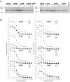
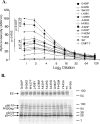
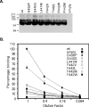
The hepatitis C virus (HCV) glycoproteins E1 and E2 form a heterodimer that mediates CD81 receptor binding and viral entry. In this study, we used site-directed mutagenesis to examine the functional role of a conserved G436WLAGLFY motif of E2. The mutants could be placed into two groups based on the ability of mature virion-incorporated E1E2 to bind the large extracellular loop (LEL) of CD81 versus the ability to mediate cellular entry of pseudotyped retroviral particles. Group 1 comprised E2 mutants where LEL binding ability largely correlated with viral entry ability, with conservative and nonconservative substitutions (W437 L/A, L438A, L441V/F, and F442A) inhibiting both functions. These data suggest that Trp-437, Leu-438, Leu-441, and Phe-442 directly interact with the LEL. Group 2 comprised E2 glycoproteins with more conservative substitutions that lacked LEL binding but retained between 20% and 60% of wild-type viral entry competence. The viral entry competence displayed by group 2 mutants was explained by residual binding by the E2 receptor binding domain to cellular full-length CD81. A subset of mutants maintained LEL binding ability in the context of intracellular E1E2 forms, but this function was largely lost in virion-incorporated glycoproteins. These data suggest that the CD81 binding site undergoes a conformational transition during glycoprotein maturation through the secretory pathway. The G436P mutant was an outlier, retaining near-wild-type levels of CD81 binding but lacking significant viral entry ability. These findings indicate that the G436WLAGLFY motif of E2 functions in CD81 binding and in pre- or post-CD81-dependent stages of viral entry.
Figures


Similar articles
-
Determinants of CD81 dimerization and interaction with hepatitis C virus glycoprotein E2.Biochem Biophys Res Commun. 2005 Mar 4;328(1):251-7. doi: 10.1016/j.bbrc.2004.12.160. Biochem Biophys Res Commun. 2005. PMID: 15670777
-
Dissecting the role of putative CD81 binding regions of E2 in mediating HCV entry: putative CD81 binding region 1 is not involved in CD81 binding.Virol J. 2008 Mar 20;5:46. doi: 10.1186/1743-422X-5-46. Virol J. 2008. PMID: 18355410 Free PMC article.
-
Expression and characterization of a minimal hepatitis C virus glycoprotein E2 core domain that retains CD81 binding.J Virol. 2007 Sep;81(17):9584-90. doi: 10.1128/JVI.02782-06. Epub 2007 Jun 20. J Virol. 2007. PMID: 17581991 Free PMC article.
-
[Role of N-linked glycans in the functions of hepatitis C virus envelope glycoproteins].Ann Biol Clin (Paris). 2007 May-Jun;65(3):237-46. Ann Biol Clin (Paris). 2007. PMID: 17502294 Review. French.
-
Topology of hepatitis C virus envelope glycoproteins.Rev Med Virol. 2003 Jul-Aug;13(4):233-41. doi: 10.1002/rmv.391. Rev Med Virol. 2003. PMID: 12820185 Review.
Cited by
-
From Structural Studies to HCV Vaccine Design.Viruses. 2021 May 4;13(5):833. doi: 10.3390/v13050833. Viruses. 2021. PMID: 34064532 Free PMC article. Review.
-
The disulfide bonds in glycoprotein E2 of hepatitis C virus reveal the tertiary organization of the molecule.PLoS Pathog. 2010 Feb 19;6(2):e1000762. doi: 10.1371/journal.ppat.1000762. PLoS Pathog. 2010. PMID: 20174556 Free PMC article.
-
Mutagenesis of the fusion peptide-like domain of hepatitis C virus E1 glycoprotein: involvement in cell fusion and virus entry.J Biomed Sci. 2009 Sep 24;16(1):89. doi: 10.1186/1423-0127-16-89. J Biomed Sci. 2009. PMID: 19778418 Free PMC article.
-
Probing the antigenicity of hepatitis C virus envelope glycoprotein complex by high-throughput mutagenesis.PLoS Pathog. 2017 Dec 18;13(12):e1006735. doi: 10.1371/journal.ppat.1006735. eCollection 2017 Dec. PLoS Pathog. 2017. PMID: 29253863 Free PMC article.
-
The neutralizing activity of anti-hepatitis C virus antibodies is modulated by specific glycans on the E2 envelope protein.J Virol. 2007 Aug;81(15):8101-11. doi: 10.1128/JVI.00127-07. Epub 2007 May 23. J Virol. 2007. PMID: 17522218 Free PMC article.
References
-
- Bartosch, B., and F. L. Cosset. 2006. Cell entry of hepatitis C virus. Virology 348:1-12. - PubMed
-
- Bartosch, B., A. Vitelli, C. Granier, C. Goujon, J. Dubuisson, S. Pascale, E. Scarselli, R. Cortese, A. Nicosia, and F. L. Cosset. 2003. Cell entry of hepatitis C virus requires a set of co-receptors that include the CD81 tetraspanin and the SR-B1 scavenger receptor. J. Biol. Chem. 278:41624-41630. - PubMed
Publication types
MeSH terms
Substances
LinkOut - more resources
Full Text Sources
Other Literature Sources

